- Home
- Users & Science
- Find a beamline
- Structural biology
- Our beamlines
- ID23-1: Gemini - Macromolecular Crystallography
- Technical Overview
- Optic Hutch Overview
- White Beam Attenuators and Beam Viewer
White Beam Attenuators and Beam Viewer
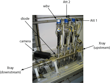
There is a total of three sets of attenuators located after the primary slits called Att 1, Att 2 and wbv (standing for White Beam viewer) when you follow the beamline dowstream. Each of them is mounted with a set of six different thickness of material and motorised in order to set automatically the required attenuation. For the sake of the thin aluminium foils it is always recommended to start by inserting in the beam the carbone attenuator before any aluminium attenuator.

The impurities present in the diamond fluoresce when they get in the white beam (essentially interacting with the low energy). A camera focuses on this position and gives an indirect and qualitative image of the shape and intensity of the white beam. Furthermore a diode recording the diffuse scattering from the pyrocarbone foil (position 0) or diamond (position 1) allows quantitative calibration of the white beam.
|
|
wbv - white beam viewer (1), C (0, 4, 5) and Al (3)
|
Att2 (Aluminium)
|
Att1 (Carbon)
|
||||||||||||||||||||
|
Position
|
0
|
1
|
2
|
3
|
4
|
5
|
|
|
0
|
1
|
2
|
3
|
4
|
5
|
|
|
0
|
1
|
2
|
3
|
4
|
5
|
|
|
Thickness in mm
|
0.5
|
Diamond
|
Empty
|
5.0
|
5.0
|
8.0
|
|
|
Empty
|
0.3
|
0.5
|
0.9
|
1.9
|
2.7
|
|
|
Empty
|
0.5
|
1.0
|
2.0
|
4.0
|
6.0
|
|


