- Home
- Users & Science
- Scientific Documentation
- ESRF Highlights
- ESRF Highlights 2001
- Magnetism and Electronic Properties of Solid
- Establishing X-ray Magnetic Linear Dichroism as a Probe of the Magnetocrystalline Anisotropy
Establishing X-ray Magnetic Linear Dichroism as a Probe of the Magnetocrystalline Anisotropy
The magnetocrystalline anisotropy energy (MAE) is the energy required to rotate the magnetisation (M) away from the easy direction of magnetisation and plays a key role in the development of magnetic storage media. In magnetic multilayers the interface MAE stabilises perpendicular magnetic anisotropy, which is important for high-storage density, and affects oscillatory exchange coupling, which governs magnetoresistance in reading devices. A microscopic understanding of the interface MAE is therefore important, but requires a probe which is element and site-specific. X-ray absorption spectroscopy (XAS) is a well-established element-specific probe of localised electronic structure since it involves transitions from core levels to states above the Fermi level. For transition metals, dipole selection rules ensure that 2p core levels are excited into the unoccupied 3d levels which are the states driving the magnetism. In recent years, X-ray magnetic circular dichroism (XMCD), which is XAS combined with circularly-polarised light, has emerged as a unique element-specific tool for separating the spin and orbital magnetic moments in ferri- and ferromagnetic systems. XMCD can then yield the orbital moment anisotropy, which is related to the MAE via a perturbation model. However, this approach is far from ideal because the orbital moment anisotropy is strictly not proportional to the MAE. Since the MAE arises from the spin-orbit interaction, a technique probing this quantity would seem far more appropriate. XAS combined with linearly-polarised light, X-ray magnetic linear dichroism (XMLD), has been proposed as a means of measuring the anisotropy in the spin-orbit interaction which can be related to the MAE using a straight forward sum rule [1]. XMLD also has the added advantage that it is sensitive to antiferromagnetism as well as ferri- and ferromagnetism.
 |
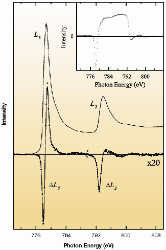
Fig. 93: Co L2,3 XAS, from 6 monolayers of Co grown on Cu( |
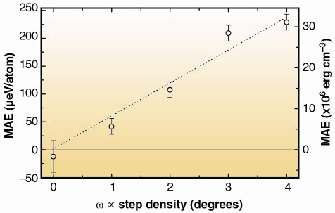
In order to verify the proposed relationship between XMLD and the MAE a Cu crystal with 5 miscut surfaces, at an angle from (001), was used as a substrate for growing ultrathin films of Co. For stepped Co surfaces, grown on Cu, it is known from surface magneto-optical Kerr measurements that the MAE increases linearly with step density [2]. The experiments were carried out on beamline ID12B (now ID8) which is an intense source of polarised soft X-rays. Co XAS spectra recorded for a stepped Co film grown on Cu (
= 4°) with M perpendicular (solid line) and parallel (broken line) to E are shown in Figure 93 along with resulting XMLD (solid circles). The double peaked structure arises due to transitions from the spin-orbit split 2p core levels to unoccupied 3d states. The inset of Figure 93 shows the integral of the XMLD which should be zero if there is no contribution from the charge anisotropy. Figure 94 shows the MAE of the stepped Co films, determined using XMLD, as a function of step density. The same linear trend for the MAE determined using XMLD proves that XMLD is directly related to the MAE. This establishes experimentally XMLD as a new element-specific tool for studying the magnetocrystalline anisotropy and opens up the possibility of, for instance, magnetic anisotropy imaging using high-resolution photoelectron emission microscopy across symmetry broken surfaces and exchange biased interfaces.
 |
Fig. 94: The Co MAE determined from the XMLD as a function of step density. The straight line is a linear fit to the data. The right axis has been calculated assuming a Co atomic volume of 8.47x10⁻²⁸ m3. |
References
[1] G. van der Laan, Phys. Rev. Lett. 82, 640 (1999).
[2] R. Kawakami et al., Phys. Rev. B 58, R5924 (1998).
Principal Publication and Authors
S.S. Dhesi (a), G. van der Laan (b), E. Dudzik (c) and A.B. Shick (d), Phys. Rev. Lett. 87, 067201 (2001).
(a) ESRF
(b) Daresbury Laboratory, Warrington (UK)
(c) Hahn-Meitner-Insitut, Berlin (Germany)
(d) University of California, Davis (USA)


