- Home
- News
- Spotlight on Science
- An X-ray bilens...
An X-ray bilens nanointerferometer
19-08-2009
A novel type of hard X-ray interferometer employing a bilens system with two parallel arrays of compound refractive lenses has been developed by scientists from France and Russia. Under coherent illumination, the bilens generates two diffraction limited mutually coherent beams. When the beams overlap they produce an interference pattern with a fringe spacing ranging from tens of nanometres to tens of micrometres. This simple way to create a X-ray standing wave in a paraxial geometry opens up the opportunity to develop new X-ray interferometry techniques to study natural and advanced man-made nanoscale materials, such as self-organised biosystems, photonic and colloidal crystals, and nanoelectronic materials.
In-line phase contrast imaging techniques have been proposed to take advantage of the brilliant and highly coherent X-ray beams provided by new synchrotron radiation sources [1]. The laser-like properties of synchrotron X-ray beams make possible the development of in-line or paraxial schemes of X-ray interferometry. One of the classical examples is the Young's double slit interferometer where interference occurs between two coherent beams created by two narrow slits [2]. However, this technique is limited in resolution to the micrometre scale by the slit size and its applicability is also limited due to significant loss of intensity.
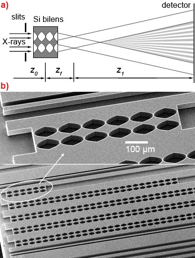
Following successful development and application of refractive optics for high energy X-rays [3-4], we propose an X-ray bilens interferometer similar to the model of the Billet split lens from classical optics. The schematic diagram of our interferometer is shown in Figure 1. It consists of two identical, parallel, planar compound refractive lenses separated transversally by a distance d. Similar to the double slit scheme, the split distance d of the bilens should be smaller than the spatial coherence length of the incoming beam defined as lcoh = λz0/S, where S is the effective source size (FWHM). As a consequence each lens is illuminated coherently, and therefore it generates a coherent, diffraction limited focal spot of size σ = λzf /Aeff , where Aeff is the absorption limited effective aperture of the lens [3]. At a distance z1 > zf d/Aeff the cones diverging from these secondary sources overlap, and interference occurs in this region of superposition. The fringe spacing or peak-to-peak width of fringes is given by Λ = λz1/d. This interference pattern has a variable period ranging from tens of nanometres to tens of micrometres, depending on the distance z1.
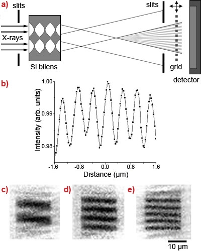
The experimental tests of the bilens interferometer were carried out at beamline ID06. Monochromatic radiation in the range of 10 – 20 keV was selected using a Si-111 double crystal monochromator. To resolve the nanometre scale interference fringes in the near field we apply scanning and imaging (Figure 2) techniques using a 0.5 µm thick Ta grid with a 400 nm periodicity (200 nm slit and 200 nm bar) behind the bilens. In the scanning mode the intensity modulations are measured with a PIN-diode by moving the grid across the fringes generated by the bilens. The Ta grid was placed at a distance of 270 mm from the bilens where the pitches of the grid and the interference pattern coincided. The vertical scan of the grid in the vertical direction is shown in Figure 2b. To visualise a nano-pattern we applied a moiré imaging technique in which two periodical patterns with slightly different pitch sizes interfere. Moving the Ta grid along the bilens optical axis, we probed the pitch of the interference pattern. At the distance of 270 mm, the grid and the interference pattern pitch coincide and Moiré fringes are absent. Depending on the grid displacement from this position the moiré pattern changes from 2 to 5 fringes with periodicity from 8 µm to 4 µm (Figures 2c-e). We emphasise that the change in the number of fringes from 4 to 5 (Figures 2d-e) corresponds to a 5 nm increase of the bilens interference pitch size and 3 mm longitudinal grid shift, demonstrating the extreme precision of the technique. A coherent moiré imaging or radiography technique using a bilens is a straightforward application. The evident advantage of moiré radiography over direct measurement of the interference fringes created by the bilens is that it greatly reduces the requirements on detector resolution while still offering submicrometre and nanometre resolution.
We have demonstrated a new and simple way to generate an X-ray periodic interference field such as a standing wave with variable period ranging from tens of nanometres to tens of micrometres. The proposed interferometer thus occupies the place between crystal and grating interferometers. The silicon bilens can generate standing waves with a 40 nm period above 50 keV. If diamond lenses were employed, then we could expect the smallest pitch to be 30 nm at energies E > 12 keV. Contrary to Bonse-Hart interferometers and X-ray standing wave techniques, the bilens interferometer generates an interference pattern without the requirement of additional optics like crystals or multilayers. The interference occurs in air at a reasonable distance from the device itself allowing great flexibility in sample size and environment.
Such coherent spatially harmonic illumination can be used for new diffraction and imaging methods to study mesoscopic materials. A phase contrast imaging technique is feasible whereby a sample is inserted into one of the beams while they are separated, as in the case of a classical interferometer. Any interaction with that beam will induce significant changes in the interference pattern, allowing the extraction of high resolution information on the sample from the new phase pattern produced. A second technique would be Moiré imaging whereby the sample is placed behind the bilens within the interference field. Standing wave techniques are evident: a sample could be scanned across the periodic interference field and secondary processes (fluorescence, secondary electrons, etc.) would be detected by a detector placed to one side of the beam.
The bilens interferometer presented here exhibits major advantages over other interferometer schemes taken from classical optics such as Young double slits, Fresnel double mirrors or bi-prisms. Manufacturing of micro-slits, mirrors and prisms for hard X-rays is a challenging technological task considering the requirements of surface and shape (edges) quality. In contrast, for silicon planar lenses, well-developed microelectronics technology is used providing superior lens quality [4]. Furthermore, unlike slits, mirrors and prisms, the bilens system can be used at high photon energies, up to 100 keV. The bilens system has the advantage over double slits as it focuses X-rays into the region where the two beams intersect, leading to an intensity gain factor for our nano-interferometer with respect to a hypothetical linear slit of 50 nm of at least 3 orders of magnitude.
In addition to imaging applications, the bilens interferometer could also be used for coherence diagnostics at existing synchrotrons. Silicon bilenses are stable under extremely powerful beams and relatively insensitive to mechanical vibrations. We expect that they will also be widely used for beam characterisation for future free electron X-ray laser sources.
References
[1] A. Snigirev et al., Rev. Sci. Instrum. 66, 5486 (1995).
[2] W. Leitenberger, S.M. Kuznetsov, A. Snigirev, Opt. Commun. 191, 91 (2001).
[3] A. Snigirev, V. Kohn, I. Snigireva, B. Lengeler, Nature 384, 49 (1996).
[4] A. Snigirev et al., SPIE 6705, 670506-1 (2007).
Principal publication and authors
A. Snigirev (a), I. Snigireva (a), V. Kohn (b), V. Yunkin (c), S. Kuznetsov (c), M. Grigoriev (c), T. Roth (a), G. Vaughan (a), C. Detlefs (a), An X-ray nanointerferometer based on Si refractive bilenses, Phys. Rev. Lett. 103, 064801 (2009).
(a) ESRF
(b) Russian Research Center 'Kurchatov Institute', Moscow (Russia)
(c) IMT RAS, Chernogolovka, Moscow region (Russia)


