Publications
|
A. Suzuki, C.S. Cheung, Y. Li, A. Hogg, P.S. Atkinson, C. Riminesi, C. Miliani, H. Liang, Time and spatially resolved VIS-NIR hyperspectral imaging as a novel monitoring tool for laser-based spectroscopy to mitigate radiation damage on paintings, Analyst (2024) |
|
The increased adoption of non-invasive laser-based techniques for analysis of cultural assets has recently called into question the non-invasiveness of the techniques in practical operation. The methods to assess the occurrence of radiation-induced alteration on paintings are very limited and none of them can predict damage. Here we present a novel multimodal imaging approach to understand the time and spatial evolution and types of laser-induced surface alterations, through simultaneous monitoring using visible and near infrared (VIS-NIR) reflectance hyperspectral imaging (HSI) and thermal imaging during Raman spectroscopy. The resultant physical and chemical changes were examined in detail by optical coherence tomography and synchrotron based micro-X-ray powder diffraction. HSI was found to be the most sensitive in detecting laser induced alternations compared with conventional methods. It is orders of magnitude more sensitive than Raman spectroscopy and even synchrotron-based micro-X-ray powder diffraction. In cases of thermally driven alterations, transient and reversible reflectance changes were found to be the first indications of laser-induced modifications and can therefore be used as precursors to prevent damage. VIS-NIR reflectance spectroscopy should be used to monitor laser-based analysis and potentially other radiation-based techniques in situ to mitigate laser induced alteration.
|
N. Palladino, M. Occelli, G. Wallez, Y. Coquinot, Q. Lemasson, L. Pichon, S. Stankic, V. Etgens & J. Salvant An analytical survey of zinc white historical and modern artists’ materials. Heritage Science 12, 47 (2024) |
|
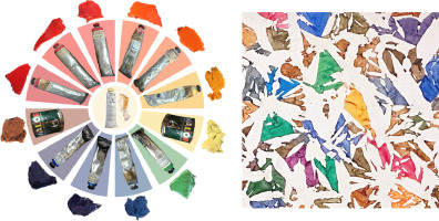
This study is the first systematic survey of a large corpus of zinc white (ZnO) artists’ materials. Zinc white is a white pigment developed within the wave of 19th-century technological developments in the paint industry. The composition, particle morphology and size, and luminescence of 49 zinc white samples from artists’ materials were characterized, including three references of known synthesis methods (indirect and direct) and synthesized by the authors (ZnO nanosmoke). The corpus included historical and modern zinc white pigment powders and paint materials from the leading European and American color manufacturers. The study aims to characterize and evaluate the variability of the properties of zinc white and its paint formulations. The reference materials presented properties in agreement with the literature: indirect ZnO exhibited submicron prismoidal blue-luminescent particles of higher purity than direct ZnO, which had larger acicular green-luminescent particles. ZnO nanosmoke presented acicular (tetrapod-like) blue/green-luminescent nanoparticles. Composition, particle morphology, size, and documentary sources suggested a production via the indirect method for the analyzed corpus. However, the luminescence behavior was more complex to interpret. The fundamental emission of ZnO was not always detected, even in pure ZnO powders. Three trends were identified: smaller ZnO particles for the most recent samples; green luminescence connected to larger particle size; fewer trace elements, and of the same type (i.e., lead, sulfur) for historical materials. Another interesting finding was the detection of hydrozincite in some powders, likely a degradation product of ZnO. In terms of methodology, cathodoluminescence proved a valuable tool for pigment identification. The study provides a database of zinc white references for pigment and artwork analysis.
|
L. Dalecky, I. Bonaduce, É. Anheim, J. La Nasa, M. L’Héronde, C. Morel, E. Catelli, S. Prati, Z. Li, L. Beck, I. Caffy, E. Delqué-Količ, A. Chevalier, L. Bertrand, "A typical postwar workshop: Insights into Simon Hantaï’s oil paint palette" Journal of Cultural Heritage, Volume 66, (2024) |
|
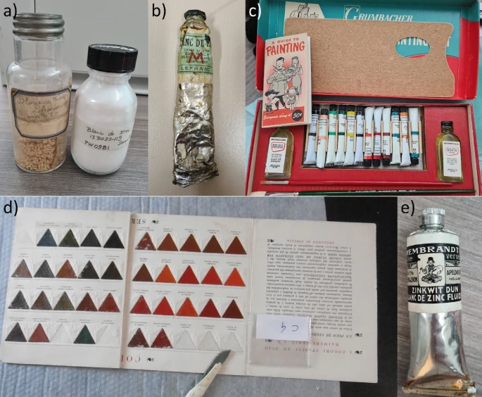
Simon Hantaï (1922–2008) was a highly influential postwar painter in Paris whose innovative serial practice, creative curiosity and theoretical convictions inspired a number of his contemporaries. His art media are typical of the period, consisting of commercial artists’ products, sold in tubes and cans, which were available to artists in Europe and beyond. We have studied a series of samples from the brands Lefebvre-Foinet, Lefranc & Bourgeois and Valor using a combination of optical and electron microscopy, accelerator-mass spectrometry carbon-14 dating, infrared spectroscopy, structural analysis, chromatographic and mass spectrometry techniques. Of particular interest is the rare access to a coherent artist’s studio collection and its dating in relation to the painter’s works. We gained precise information on paint formulations, including main binders and pigments, as well as additives, such as free metal soaps, beeswax and pine resin. This suggests the value of further research into paint formulations and their identification in paintings from the second half of the 20th century. These materials were studied for their capacity as possible references for future analysis of the painter’s artworks. The high degree of hydrolysis of the oil binder and alteration, notably by saponification, leads us to question the significance of these materials and the handling of the data generated towards comparative studies. These samples have a history; considering them as pristine references for comparative studies with the works of artists of the period cannot be done at the expense of their own materiality — and in particular their physico-chemical evolution over time in their specific environment
|
|
|
 G. Pastorelli, A. S. Ortiz Miranda, E. Avranovich Clerici, P. d'Imporzano, B. Vinther Hansen, K. Janssens, G. R. Davies, N. Borring, "Darkening of lead white in old master drawings and historic prints: A multi-analytical investigation", Microchemical Journal, (2024).
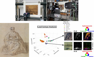
Old master drawings and historic prints often feature white highlights, which are typically painted using lead white, one of the most widely used historical white pigments. However, it has been observed that many of these highlights discolour over time, becoming dark brown or black due to unclear degradation processes. This phenomenon not only misrepresents the original artefacts, threatening their suitability for public display, but also diminishes their longevity. To ensure their preservation, it is essential to determine why some lead white highlights in these museum objects retain their light tones while others are prone to darkening. The objective of this study was to identify the relationships between the composition, provenance, and production methods of lead white pigments, and their role in the discolouration observed on drawings, lithographs and early photographs. Selected samples and artefacts were examined using a range of analytical techniques, namely X-ray fluorescence spectroscopy (XRF), X-ray powder diffraction (XRPD), and lead isotope analysis. While XRF analyses confirmed the presence of lead as the primary element in the majority of the highlights, XRPD measurements identified a variety of lead compounds such as the carbonates cerussite and hydrocerussite alongside galena—a black crystalline sulfide—and lead sulfates. Additionally, isotope analyses classified the lead raw materials into five main groups. Through these measurements, the examined lead white pigments were categorised based on their compositional properties in relation to the raw materials used, as well as their geographical and temporal origin. A significant finding is that lead white pigments from different production periods, spanning from older to more modern, may be characterised by varying proneness to discolouration irrespective of their provenance.
|
C. de Mecquenem, M. Eveno, M. Alfeld et al. “A multimodal study of smalt preservation and degradation on the painting “Woman doing a Libation or Artemisia” from an anonymous painter of the Fontainebleau School” Eur. Phys. J. Plus 138, 185 (2023).
|
|
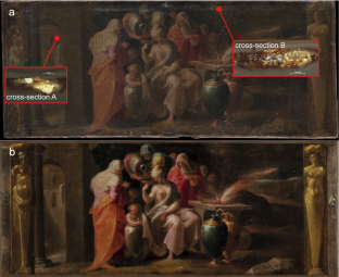
The blue pigment smalt, a synthetic potash glass tinted with cobalt, was widely used between the sixteenth and the eighteenth centuries. As part of a study on the alteration of smalt and the reconstitution of its original color, the painting: Woman doing a Libation or Artemisia (Fontainebleau school, 1570) was examined in which the artist used smalt as a blue pigment, which is now degraded. Noninvasive imaging was performed using macro-2D X-ray fluorescence and reflectance imaging spectroscopy to get an overview of the artist’s palette and its distribution. Samples prepared as cross sections were also analyzed by scanning electron microscopy coupled with energy dispersive spectroscopy, micro-X-ray absorption near-edge structure spectroscopy and synchrotron micro-X-ray diffraction imaging to determine the preservation state of the smalt as well as structural information on other pigments adjacent to smalt grains in individual paint layers, which could play a role in the degradation process. On the one hand, the study conducted on the alteration of smalt has shown that it is very weathered and mixed with hydrocerussite, which could be a factor that would facilitate the alteration. On the other hand, these analyses have made it possible to identify and locate the pigments used, which will be the basis for the virtual reconstruction of the color of the painting.
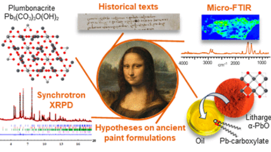 |
V. Gonzalez, G. Wallez, E. Ravaud, M. Eveno, I. Fazlic, T. Fabris, A. Nevin, T. Calligaro, M. Menu, V. Delieuvin, and M. Cotte, "X-ray and Infrared Microanalyses of Mona Lisa’s Ground Layer and Significance Regarding Leonardo da Vinci’s Palette", Journal of the American Chemical Society, Article ASAP, (2023)
|
An exceptional microsample from the ground layer of Leonardo da Vinci’s Mona Lisa was analyzed by high-angular resolution synchrotron X-ray diffraction and micro Fourier transform infrared spectroscopy, revealing a singular mixture of strongly saponified oil with high lead content and a cerussite (PbCO3)-depleted lead white pigment. The most remarkable signature in the sample is the presence of plumbonacrite (Pb5(CO3)3O(OH)2), a rare compound that is stable only in an alkaline environment. Leonardo probably endeavored to prepare a thick paint suitable for covering the wooden panel of the Mona Lisa by treating the oil with a high load of lead II oxide, PbO. The review of Leonardo’s manuscripts (original and latter translation) to track the mention of PbO gives ambiguous information. Conversely, the analysis of fragments from the Last Supper confirms that not only PbO was part of Leonardo’s palette, through the detection of both litharge (α-PbO) and massicot (β-PbO) but also plumbonacrite and shannonite (Pb2OCO3), the latter phase being detected for the first time in a historical painting.
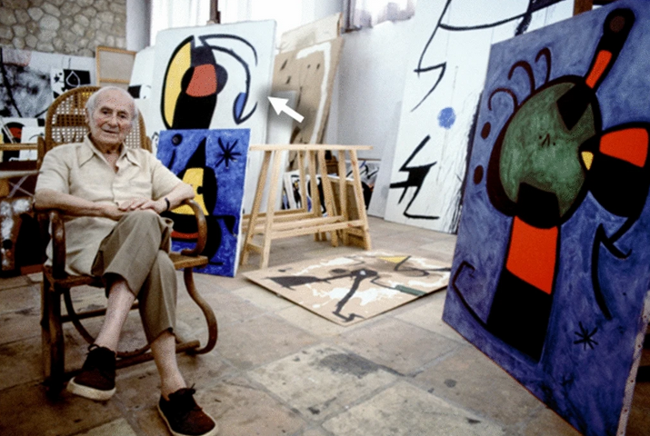 |
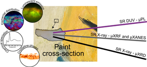
M. Gomez Lobon, M. Ghirardello, E. Juncosa Darder, C Palomino Cabello, M. Bauza, M.Cotte, A. Burnstock, A. Nevin, S. Rita Amato, F. Caterina Izzo and D. Comelli, "A study of cadmium yellow paints from Joan Miró’s paintings and studio materials preserved at the Fundació Miró Mallorca", Herit Sci 11, 145 (2023).
|
The deterioration of cadmium yellow paints in artworks by Joan Miró (1893–1983) and in painting materials from his studios in Mallorca (Spain) was investigated. Analysis of samples from Miró’s paintings and from paint tubes and palettes showed that degraded paints are composed of poorly crystalline cadmium sulfide/zinc cadmium sulfide (CdS/Cd1−xZnxS) with a low percentage of zinc, in an oil binding medium. Cadmium sulfates were identified as the main deterioration products, forming superficial white crusts detected using SR µXANES and µXRD techniques. Time-resolved photoluminescence measurements demonstrated that highly degraded samples display a pink/orange emission from the paint surface with a microsecond lifetime, a phenomenon observed in other degraded cadmium yellow paints. In agreement with recent studies on altered cadmium paints, these results suggest that the stability of the paint is related to its manufacturing method, which affects the degree of crystallinity of the resulting pigment. This, together with the environmental conditions in which artworks have been exposed, have induced the degradation of yellow paints in Miró’s artworks. It was finally noted that the paints exhibiting alteration in the analysed Miró artworks have a chemical composition that is very similar to the tube paint ‘Cadmium Yellow Lemon No. 1’ produced by Lucien Lefebvre-Foinet. Indeed, paint tubes from this brand were found in the studio, linking the use of this product with Miro’s degraded artworks.
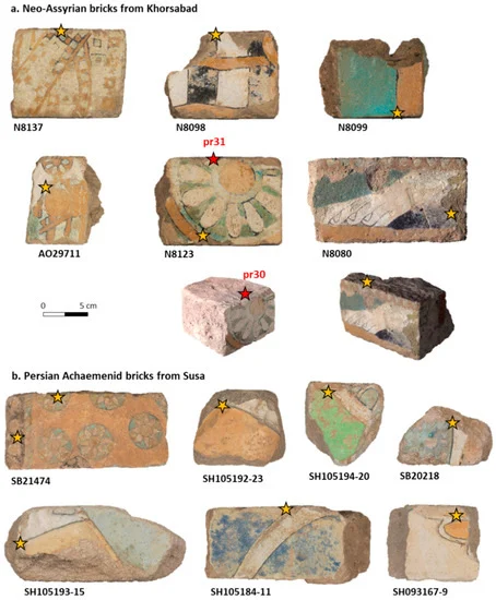 |
E. Beauvoit, A. Bouquillon, O. Majérus, D. Caurant, J. Cuny, A. Thomas, “Comparative Study of Architectural Bricks from Khorsabad and Susa Sites: Characterization of Black Glazes”, Heritage, (2023). |
In this study, the well-preserved glazes of 13 colored bricks representative of the decoration of the palaces of Sargon II (Khorsabad, 8th century BC) and of Darius I (Susa, 6th century BC) were examined. The purpose of this research is to gather information about the ancient brick manufacturing processes by examining the colored glazes and, in particular, black glazes using a combination of methods that included optical microscopy, SEM-EDX, synchrotron µ-XRD, and µ-Raman spectroscopy. The results revealed different coloring techniques for producing black glazes in the Neo-Assyrian and Persian Achaemenid periods. Regarding the black glazes of Susa, it is particularly interesting to note that their chemical composition varies according to the function of the glazes on the bricks: manganese oxide (for colored fields of glaze) and iron-rich compounds (for raised lines separating glazed areas). In comparison, the black glazes from Khorsabad are characterized by the presence of spherical copper sulfide and galena nanoparticles (ranging from less than 100 nm to about 1 µm) for both the glazed areas and the separating lines. This coloring technique to obtain black glazes is very rarely described in the literature, as well as the mechanism of formation of these spherical nanoparticles.
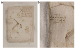 |
N. Guarnieri, M. Ghirardello, S. Goidanich, D.Comelli, D. Dellasega, M. Cotte, E. Fontana and L. Toniolo, “Imaging and micro‑invasive analyses of black stains on the passepartout of Codex Atlanticus Folio 843 by Leonardo da Vinci”, Scientific Reports, (2023). |
XRD micro-analyses (ID13 and ID22, through the historical materials BAG) and µXANES analyses (ID21) have been combined with laboratory imaging techniques to identify the nature of black stains present on the passepartout close to the margins of Folio 843 of Leonardo da Vinci’s Codex Atlanticus. Instead of the microbiological deterioration which was formerly proposed, black nano-particles of metacinnabar (β‑HgS) have been identified as the component of the black staining.
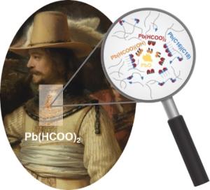 |
V. Gonzalez, I. Fazlic, M. Cotte, F. Vanmeert, A. Gestels, S. De Meyer, F. Broers, J. Hermans, A. van Loon, K. Janssens, P. Noble, K. Keune, “Lead(II) Formate in Rembrandt's Night Watch: Detection and Distribution from the Macro- to the Micro-scale”, Angew. Chem. Int. Ed. (2023). |
X-ray powder diffraction mapping at multiple length scales revealed the unusual presence of lead(II) formate, Pb(HCOO)2, in several areas of The Night Watch, Rembrandt's most famous painting. A possible chemical pathway resulting in the formation of this compound in historical oil paint was explored via micro-analysis, notably using synchrotron radiation. New clues on the reactivity of metallic driers in oil systems were thus gathered.
|
|
|
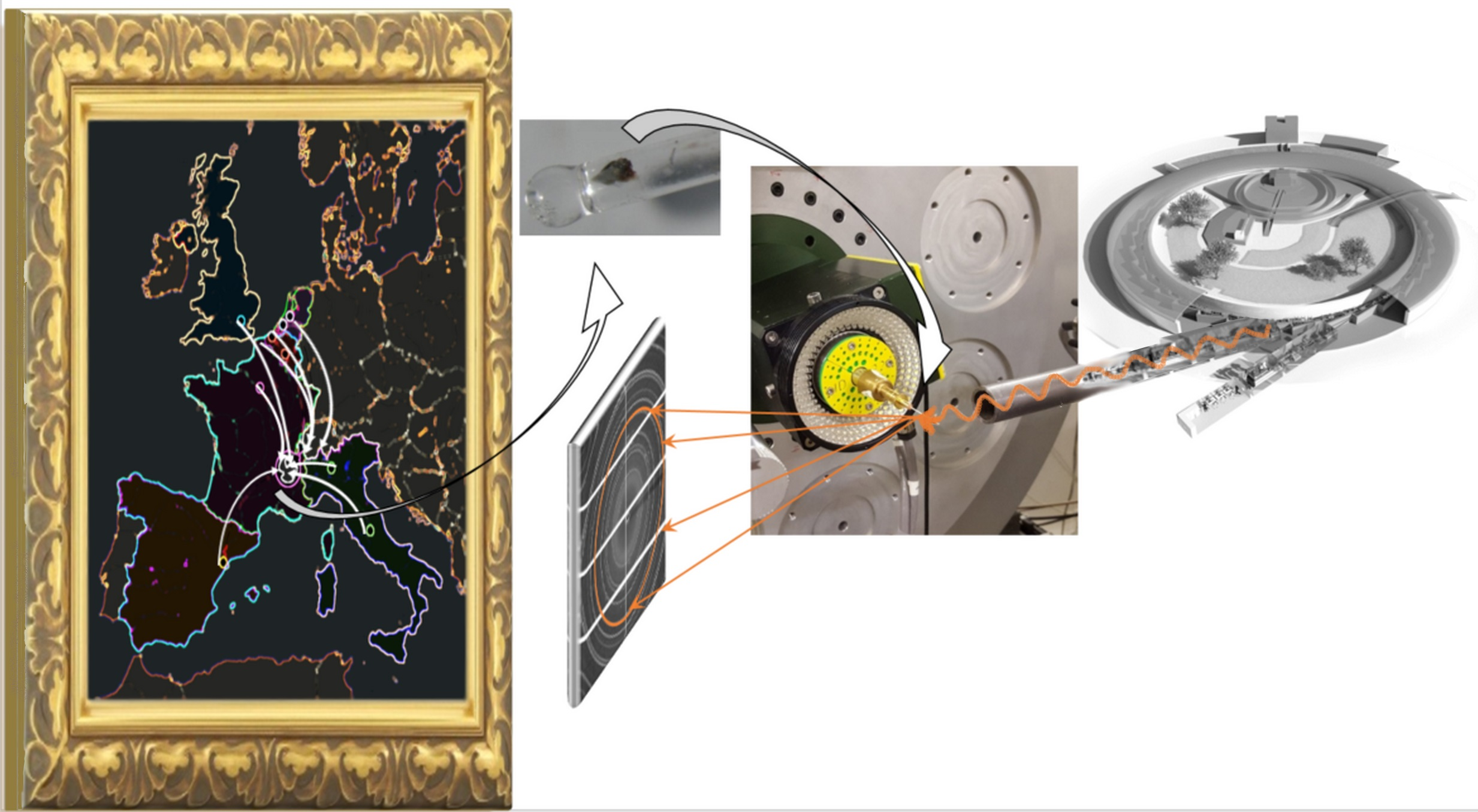 Cotte M, Gonzalez V, Vanmeert F, Monico L, Dejoie C, Burghammer M, Huder L, de Nolf W, Fisher S, Fazlic I, Chauffeton C, Wallez G, Jiménez N, Albert-Tortosa F, Salvadó N, Possenti E, Colombo C, Ghirardello M, Comelli D, Avranovich Clerici E, Vivani R, Romani A, Costantino C, Janssens K, Taniguchi Y, McCarthy J, Reichert H, Susini J. The “Historical Materials BAG”: A New Facilitated Access to Synchrotron X-ray Diffraction Analyses for Cultural Heritage Materials at the European Synchrotron Radiation Facility. Molecules (2022)
The European Synchrotron Radiation Facility (ESRF) has recently commissioned the new Extremely Brilliant Source (EBS). The gain in brightness as well as the continuous development of beamline instruments boosts the beamline performances, in particular in terms of accelerated data acquisition. This has motivated the development of new access modes as an alternative to standard proposals for access to beamtime, in particular via the “block allocation group” (BAG) mode. Here, we present the recently implemented “historical materials BAG”: a community proposal giving to 10 European institutes the opportunity for guaranteed beamtime at two X-ray powder diffraction (XRPD) beamlines—ID13, for 2D high lateral resolution XRPD mapping, and ID22 for high angular resolution XRPD bulk analyses—with a particular focus on applications to cultural heritage. The capabilities offered by these instruments, the specific hardware and software developments to facilitate and speed-up data acquisition and data processing are detailed, and the first results from this new access are illustrated with recent applications to pigments, paintings, ceramics and wood.
|
N. Oriols, N. Salvadó, T. Pradell, N. Jiménez, M. Cotte, V. Gonzalez, S. Butí, "Carbonation of fresco mural paintings with a dolomitic mortar", Cement and Concrete Research, Volume 157 (2022). |
|
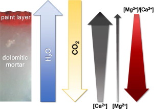
An innovative approach involving a collection of experiments to mimic and assess the different conditions (at selected time-pH-[Mg2+]-humidity) existing during the carbonation process in dolomitic mortars is proposed with a combination of micro-analytical imaging techniques, namely: optical and electron microscopy, micro-Fourier-Transform Infrared spectroscopy, Raman spectroscopy and synchrotron-based micro X-ray diffraction. The carbonation process was studied from short time scale to long time scale and from simple aqueous saturated solutions to fresco mock-up samples. These techniques allowed identifying both crystallized and amorphous components and revealing their in-depth distribution in multi-layered systems. The presence of Mg2+ influences the microstructure and composition of dolomitic mortars, resulting in the presence of aragonite concurrent to calcite. In later stages, i.e. once Ca2+ has transformed into carbonate, brucite can lead to the formation of magnesium carbonates and hydroxycarbonates. The results contribute explaining the lability of old dolomitic mortars observed in historical fresco paintings.
|
M. Ghirardello, V. Gonzalez, L. Monico, A.Nevin, D. MacLennan, C. Patterson, M. Burghammer, M. Réfrégiers, D. Comelli, M. Cotte, "Application of Synchrotron Radiation-Based Micro-Analysis on Cadmium Yellows in Pablo Picasso's Femme", Microscopy and Microanalysis, 1-10 (2022). |
|
The cultural heritage community is increasingly exploring synchrotron radiation (SR) based techniques for the study of art and archaeological objects. When considering heterogeneous and complex micro-samples, such as those from paintings, the combination of different SR X-ray techniques is often exploited to overcome the intrinsic limitations and sensitivity of the single technique. Less frequently, SR X-ray analyses are combined with SR micro-photoluminescence or micro-Fourier Transform Infrared spectroscopy, which provide complementary information on the molecular composition, offering a unique integrated analysis approach. Although the spatial correlation between the maps obtained with different techniques is not straightforward due to the different volumes probed by each method, the combination of the information provides a greater understanding and insight into the paint chemistry. In this work, we discuss the advantages and disadvantages of the combination of X-ray techniques and SR-based photoluminescence through the study of two paint micro-samples taken from Pablo Picasso's Femme (1907). The painting contains two cadmium yellow paints (based on CdS): one relatively intact and one visibly degraded. SR micro-analyses demonstrated that the two Cd-yellow paints differ in terms of structure, chemical composition, and photoluminescence properties. In particular, on the basis of the combination of different SR measurements, we hypothesize that the degraded yellow is based on nanocrystalline CdS with high presence of Cd(OH)Cl. These two characteristics have enhanced the reactivity of the paint and strongly influenced its stability.
|
M. Cotte, K. DollMan, V. FernanDez, V. Gonzalez, F. VanMeert, L. Monico, C. Dejoie, M. Burghammer, L. Huder, S. Fisher, W. De Nolf, I. Fazlic, H. Castillo-Michel, M. Salomé, M. Ghirardello, D. Comelli, O. Mathon and P. Tafforeau, "New Opportunities Offered by the ESRF to the Cultural and Natural Heritage Communities" Synchrotron Radiation News 3 5:5, pages 3-9. Communities (2022) |
|
partners


European Synchrotron Radiation Facility - 71, avenue des Martyrs, CS 40220, 38043 Grenoble Cedex 9, France.