Overview
BM18 is dedicated to high sensitivity phase-contrast tomography in large and complex samples.
The main techniques used on the beamline are hierarchical phase-contrast tomography and propagation phase-contrast imaging. BM18 has a high automation level for high throughput.
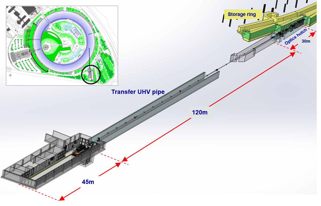
BM18 is one of the ESRF's long beamlines, measuring 220 metres, with up to 36 metres for propagation phase-contrast. The Experimental hutch measures 45 metres in length and uses a large polychromatic beam at high energy (50-280 keV).
Currently, the maximum sample size is 30kg, 30 cm diametre and 50 cm vertical field of view.
In the future, the beamline will be able to scan samples of up to 2.5m and 300 kg.
Construction of BM18 started in 2018 and the beamline opened in User Service Mode in 2022. It is expected to reach its full technical capabilities by the end of 2024.
Improvements from refurbishment and EBS
- BM18 has currently the smallest X-ray source for a large beam at high energy worldwide.
- The 35cm beam has the highest coherence worldwide for high-energy X-ray imaging.
- A large resolution range: 0.7-80 µm
Areas of research
|
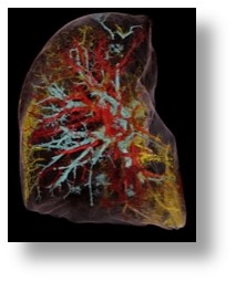
Biomedical Imaging
|
|
|
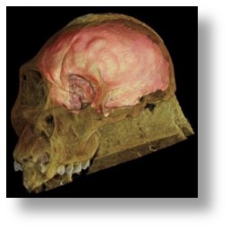
Natural and Cultural Heritage
|
|
|
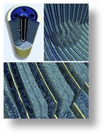
Material Sciences
|
|
|
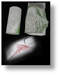
Geology
|
|
|
Industrial applications
|
|
A (very) long beamline
 |
|
The position of BM18 in relation to the ESRF storage ring and central building. |
Video resources
| Example of industrial part (composite T-bracket) imaged in multiscale approach at 42, 15, 4 and 0.65µm on BM18. Test experiment performed during the first month of beamtime on BM18 in collaboration with Fraunhofer EZRT. | |
| Effects of covid19 disease on a upper lung lobe of a deceased patient in 2019. Video shows the strong vascular damages as well as airtrack damage and the strong effect on the alveolae. The scans used for this movie at 25, 6.5µm and 2.5µm were performed on BM05 in collaboration with the UCL and lead to the development of the Hierarchical Phase-Constrat Tomography (HiP-CT) that is the base of the Human Organ Atlas project that is now running on BM18. | |
| Illustration of the Human Organ Atlas with a composite showing all the organs of a single body donor put back in place in a skeleton model. The HiP-CT is able to image complete organs, and then zoom-in anywhere in these organs down to histological resolution in 3D. More information can be found on the Human organ Atlas Hub webpage. | |
| 3D rendering of the BM18 experimental hutch during its conception phase. It shows the size of the future large sample stage compared to the present one. The large detector stage installed in the hutch is not integrated in this movie. | |
| Example of a fossil arthropod inclusion in an amber block imaged on BM18 during its initial test phase. |
