- Home
- Users & Science
- Find a beamline
- X-ray nanoprobe
- ID21 - X-ray Microscopy Beamline
- Scanning x-ray microscopes (nano-SXM and SXM-II)
Scanning x-ray microscopes (nano-SXM and SXM-II)
The two Scanning X-ray Microscopes (nano-SXM and SXM-II)
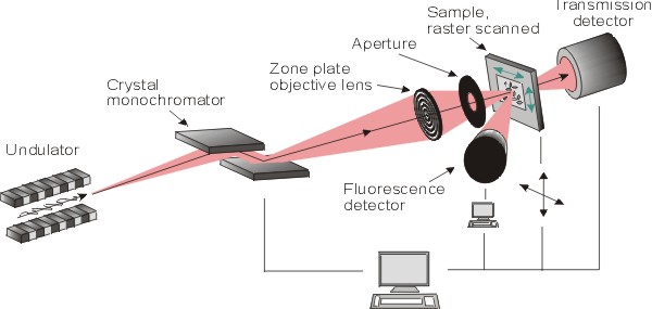
The ID21 beamline is dedicated to micro and nano-X ray spectroscopy, 2D X-ray fluorescence (XRF) mapping and X-ray absorption spectroscopy (XAS), in the tender X-ray domain (2.1-10.5keV). These two techniques can be combined to acquire multi-energy XRF maps. In brief, this beamline offers 2D elemental mapping and speciation information (in 0D, 1D and 2D), and is particularly optimized for the detection and chemical analysis of elements from P to Zn. Heavier elements can be analyzed through their L and M absorption edges. Two scanning X-ray microscopes are available for micro and nano-analyses.
[ Source characteristics] [ SXM II end-station] [ SXM-II sample preparation] [ Nano SXM sample preparation]
The beamline has recently been refurbished, and offers now two scanning X-ray microscopes: a new nanoscope, "nano-SXM", optimized for nano-analysis, which complements and outperforms the "old" microscope preserved for the study of "large" samples ("SXM-II").
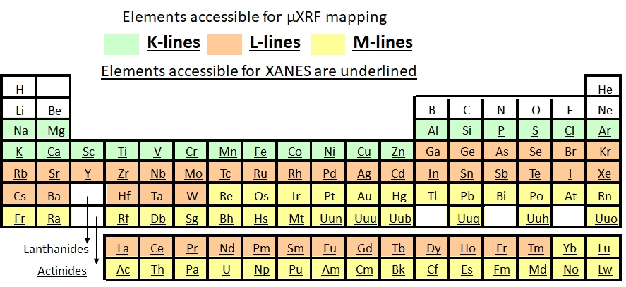
Both microscopes are operated under vacuum, either at room temperature or in cryogenics conditions. The X-ray energy range goes from 2.1 to 10.5 keV, thus giving access to the K-edges of Phosphorus to Zinc, and to the L- and M-edges of some heavier elements for micro-fluorescence and micro-XANES measurements. Please consult the following periodic table for further details.
The signal is mainly measured in X-ray fluorescence mode (with SDD detectors) but it is also possible to measure transmission signal.
The beam flux of the order of 1010- 1011 photons/s/Si<111> bandwidth.
The new X-ray nanoscope (nano-SXM) is optimized for nano-analyses. The beam can be focused down to <150nm, using either a Pt-coated (for high energy) or a Ni-coated (for low energy) KB focusing system (still under commissionning). The sample is orthogonal to the beam, and XRF is detected on both sides of the sample. Different sample holders are available.
The "old" one (SXM-II) can be operated in unfocused mode (beam defined by slits from 300 to ~50µm) and with a Ni-coated Kirkpatrick-Baez mirrors focusing system (beam down to ~0.3um ver x0.7um hor). This KB system covers all energies except the Ni K-edge. The sample is oriented with an angle of 62degrees, with respect to the incident beam, and the XRF is collected by a single SDD detector. Various sample holders are available.
Upstream the microscopes, a fixed exit double crystal monochromator equipped with Si<111>, Si<311> crystals is used to select and/or scan the energy of the x-ray beam.
partners


European Synchrotron Radiation Facility - 71, avenue des Martyrs, CS 40220, 38043 Grenoble Cedex 9, France.