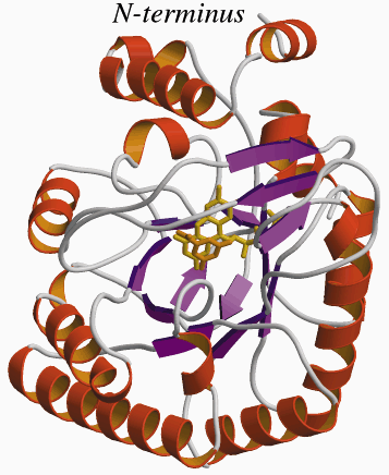
Figure 17
Fig. 17: The structure of the Escherichia coli dihydroorotate dehydrogenase. The protein forms an /ß-barrel with two extra helixes at the N-terminal lying on the side of the barrel, when compared with other DHOD structures. These helixes are proposed to be the membrane associating part of the enzyme. The FMN group and the product orotate are represented as stick models (FMN, yellow; orotate, orange).
| back to: Dihydroorotate Dehydrogenase from Escherichia coli |


