- Home
- Users & Science
- Scientific Documentation
- ESRF Highlights
- ESRF Highlights 2005
- Structural Biology
- X-ray Crystallography Study on Ribosome Recycling: Structural Explanation for RRF-mediated Subunit Dissociation
X-ray Crystallography Study on Ribosome Recycling: Structural Explanation for RRF-mediated Subunit Dissociation
Ribosome recycling is commonly referred to as the fourth stage of translation as it follows the initiation, elongation and termination phases. Ribosomes entering the recycling phase contain an mRNA with a stop codon positioned at the A site, and an uncharged (deacylated) tRNA at the P site. In bacteria, this functional state is recognised by the ribosome recycling factor (RRF), which together with elongation factor G (EF-G) and initiation factor IF3, promotes both the dissociation of the ribosome into subunits and the release of the mRNA and tRNA, thus recycling the components for the next round of translation. The importance of ribosome recycling is emphasised by the universal presence of RRF in bacteria, as well as in the mitochondria and chloroplasts, but not in the cytoplasm, of eukaryotic cells. Deletion of frr, the gene encoding RRF, is lethal in all the bacteria tested so far. Moreover, in Mycoplasma species, which have severely reduced genome sizes due to removal of nonessential genes, frr has been retained. The cellular importance and kingdom distribution of RRF thus make bacterial ribosome recycling an attractive target for drug design, a prerequisite of which is a high-resolution structure of the ribosomal binding site of RRF.
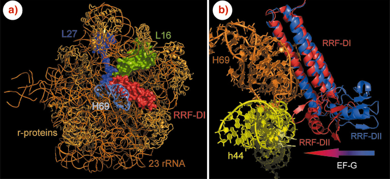
Using data collected on ID29, we have determined the X-ray structure of the ribosome binding domain of RRF (RRF-DI) bound to the large ribosomal subunit of the eubacterium Deinococcus radiodurans at 3.3 Å resolution (Figure 84a). Our study confirms the general conclusions observed in lower resolution (~12 Å) cryo-EM studies [2]. However, the position of RRF found in the cryo-EM analysis must be rotated 7° and shifted by 8 Å to be aligned with the position determined here, thus significantly altering the predicted sites of interaction. In addition, we observe multiple contacts that were not seen in the previous studies. In our study, the atomic details of the interaction of RRF with the large ribosomal subunit reveal that domain I of RRF contacts (exclusively) elements involved with tRNA binding and/or translocation (Figure 84a): (i) nucleotides G2252-G2254 of the P loop (H80), which play an important role for the positioning of the tRNA in the P-site, (ii) the base of A2602 present in H93, which has been suggested to guide the CCA-ends of the tRNA from the A- to the P-site during translocation, and (iii) the ribosomal proteins L16 and L27 at the peptidyl-transferase centre, which have been implicated in positioning of tRNAs at the P site.
 |
|
Fig. 84: (a) RRF-DI (red) interacts with helix 69 (H69) and ribosomal proteins L16 (green) and L27 (magenta) on the D. radiodurans 50S subunit. (b) EF-G binding induces a shift in the position (as indicated by arrow) of RRF-DII towards h44 of the 30S subunit leading to disruption of the intersubunit bridge contact between h44 (yellow) and H69 (orange). |
The most extensive contacts between RRF-DI and the 50S subunit are with helices 69 and 71 (H69–H71) of domain IV of the 23S rRNA (Figure 84a and b). In the 70S ribosome, H69 and H71, make contact with h44 of the 30S subunit, to form stable inter-subunit bridges [2]. Binding of RRF-DI to the 50S induces movement of H69 away from the stalk region, to resemble more closely the position observed in the Thermus thermophilus 70S ribosome [2]. However, the loop of H69 in the D50S-RRF-DI structure has a different and more open conformation compared to that in the 70S, such that its tip is shifted by 20 Å towards h44 of the small subunit.
To understand how RRF and EF-G interact on the ribosome during recycling we modeled a RRF-EFG-70S complex by (i) superimposing domain I of known unbound RRF structures with the position of RRF-DI on the 50S subunit, (ii) modeling a 70S-RRF structure by combining the high resolution 30S subunit with our 50S structure on the basis of the known 70S ribosome structure [2], and (iii) docking EF-G on the ribosome on the basis of 70S-EFG cryo-EM reconstructions. The resulting model suggests that domain III and IV of EF-G predominantly make contact with domain II of RRF (RRF-DII) upon binding. Cohabitation requires RRF-DII to move towards h44 on the 30S subunit (indicated by arrow in Figure 84b). This leads us to suggest that splitting of the 70S ribosomes into subunits during recycling results from disruption of the universally conserved intersubunit bridges through a combination of the action of RRF-DI on H69 and the EF-G induced action of RRF-DII on h44. Support for this model has been born out by recent cryo-EM structures of EF-G and RRF on the large ribosomal subunit [3].
References
[1] R.K. Agrawal et al., Proc. Natl Acad. Sci. USA 101, 8900–8905 (2004).
[2] M.M. Yusupov et al., Science 292, 883–896 (2001).
[3] N. Gao et al., Mol Cell. 18, 663-74 (2005).
Principal Publication and Authors
D.N. Wilson (a), F. Schluenzen (a), J.M. Harms (a), T. Yoshida (b), T. Ohkubo (b), R. Albrecht (a), J. Buerger (a), Y. Kobayashi (b), P. Fucini (a) EMBO J. 24, 251-260 (2005).
(a) Max-Planck Institute for Molecular Genetics, Berlin (Germany)
(b) Graduate School of Pharmaceutical Sciences, Osaka University (Japan)


