- Home
- Users & Science
- Scientific Documentation
- ESRF Highlights
- ESRF Highlights 2011
- X-ray imaging
- Local structure of single Co-implanted ZnO nanowires investigated by nano-X-ray absorption spectroscopy
Local structure of single Co-implanted ZnO nanowires investigated by nano-X-ray absorption spectroscopy
Cobalt-doped zinc oxide nanowires offer unique advantages for spintronics applications owing to their large geometrical aspect ratio and predicted room-temperature ferromagnetism [1]. However, controlled doping of semiconductor nanowires with transition metals during growth remains a challenge. Ion implantation is an alternative that allows a better control of both concentration and distribution of transition metals ions. Determining the doping homogeneity and local order of individual nanostructures is crucial to understand their behaviour in nanodevices. The average local atomic structure and secondary phases in ensembles of nanowires have already been studied by X-ray absorption near edge structure (XANES) and extended X-ray absorption fine structure (EXAFS) [2]. However, the examination of single nanowires remains an experimental challenge mainly due to the beam instability during the energy scan, and chromaticity of the nanofocusing lens. In this work, we have used a 100x100 nm2 monochromatic X-ray beam at station ID22NI to explore the short-range order in single Co implanted ZnO nanowires. In addition, the linear polarisation of the synchrotron nanobeam made our study capable of detecting preferentially oriented defects induced by the ion implantation process.
 |
|
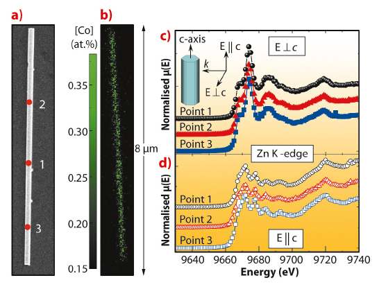
Fig. 135: a) SEM image of a single Co implanted nanowire. b) Elemental map collected at 12 keV for Co with the respective atomic fraction estimated from the XRF quantification. Zn K edge XANES spectra recorded along the nanowire: c) with the c-axis oriented perpendicular, and d) parallel to the electric field vector of the X-ray nanobeam. |
Figure 135a shows the scanning electron microscopy (SEM) image of a single nanowire. The positions indicated by 1, 2, and 3 correspond to the areas where XANES and EXAFS spectra were recorded. The elemental map of Co with the respective concentration estimated from the XRF quantification is displayed in Figure 135b, showing homogeneous distributions of Co along the nanowire without any signature of clusters or nanoaggregates. XANES data from the nanowire with its c-axis oriented perpendicular and parallel to the polarisation vector of the X-ray beam are shown in Figures 135c and 135d, respectively. The spectra exhibit the peaks associated to the hexagonal structure, without any evidence of lattice damage in the nanowire.
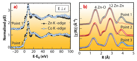
The chemical state of the implanted Co ions was investigated by nano-XANES. Figure 136a shows the XANES spectra around the Zn K edge (solid circles) and Co K edge (open circles) taken at points 1 and 2. Despite the low Co content, the quality of the XANES data around the Co K edge is good and reproduces well the oscillations of the Zn K edge spectra at both points, suggesting Co ions incorporated into the wurtzite host lattice on the Zn sites. Furthermore, the good match between the XANES spectra of the nanowire and that of a high quality wurtzite Zn0.9Co0.1O epitaxial film [3] (not shown), suggests oxidation state 2+ for implanted Co ions. Finally, nano-EXAFS measurements around the Zn K-edge allow us to study the local order of the host lattice. Figure 136b shows the magnitude of the Fourier transforms of the EXAFS functions. The spectra show two dominant peaks related to the first O- and second Zn-shells. In general, there is no evidence of amorphisation, and the Zn-O and Zn-Zn spacings remain equal to those of pure ZnO (1.98 and 3.25 Å) along the nanowire. This confirms the good recovery of the radiation damaged ZnO lattice through the thermal annealing.
 |
|
Fig. 136: a) XANES spectra around the Zn K edge (solid circles) and Co K edge (open circles) taken at points 1 and 2. For comparison between Zn and Co XANES spectra, the energy has been rescaled to the respective absorption K edges calculated from the first derivative of the XANES signal. b) Magnitude of the Fourier transforms of the EXAFS functions (open symbols) and their best fits (solid lines). |
In summary, we have carried out X-ray absorption spectroscopy in different regions along single nanowires. Our findings revealed implanted ions incorporated homogeneously along the nanowire as Co2+ and occupied Zn sites into the host lattice. The radiation damage in the ZnO host lattice was completely recovered through thermal annealing.
Principal publication and authors
J. Segura-Ruiz (a), G. Martinez-Criado (a), M.H. Chu (a), S. Geburt (b) and C. Ronning (b), Nano Lett. 11, 5322-5326 (2011).
(a) ESRF
(b) Institute of Solid State Physics, University of Jena (Germany)
References
[1] Y.W. Heo, D. Norton, L. Tien, Y. Kwon, B. Kang, F. Ren, S. Pearton and J. LaRoche, Mater. Sci. Eng. R 47, 1–47 (2004).
[2] B.D. Yuhas, S. Fakra, M.A. Marcus and P. Yang, Nano Lett. 7, 905–909 (2007).
[3] A. Ney, K. Ollefs, S. Ye, T. Kammermeier, V. Ney, T.C. Kaspar, S.A. Chambers, F. Wilhelm and A. Rogalev, Phys. Rev. Lett. 100, 157201 (2008).


