- Home
- Users & Science
- Scientific Documentation
- ESRF Highlights
- ESRF Highlights 2011
- Soft condensed matter
- Promiscuous particles caught in the act
Promiscuous particles caught in the act
Colloidal nanocomposites are currently of great interest for both fundamental studies and practical applications. Recently, we reported a route to well-defined core-shell polymer-silica nanocomposite particles based on the physical adsorption (or heteroflocculation) of 20 nm silica nanoparticles onto relatively large sterically-stabilised poly(2-vinylpyridine) (P2VP) latex particles in aqueous solution [1]. These hybrid organic-inorganic particles exhibit a fascinating phenomenon: addition of excess P2VP latex particles to a colloidal dispersion of P2VP-silica nanocomposite particles leads to the spontaneous redistribution of the silica nanoparticles such that partial coverage of all the latex particles is achieved [2]. In this study, time-resolved small-angle X-ray scattering (SAXS) measurements undertaken at station ID02 allowed the kinetics of both silica adsorption and silica redistribution onto the latex to be monitored on very short timescales.
Rapid mixing of two solutions can be achieved with minimal dead time by using a stopped-flow apparatus, thus allowing the early stages of a rapid self-assembly process to be studied with millisecond time resolution. Moreover, the relatively high electron density contrast between the latex core and the silica shell makes SAXS well-suited for time-resolved measurements of the core-shell particle formation in situ. A stroboscopic approach was adopted to achieve a resolution of 0.01 seconds.
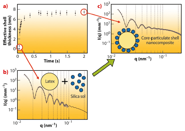
Time-resolved SAXS confirmed that the silica nanoparticles adsorb onto a 461 nm diameter sterically-stabilised P2VP latex to form a complete monolayer shell approximately 2.0 seconds after mixing the latex and silica dispersions (Figure 56). The lower time limit of our experimental setup is just sufficient to observe the binary mixture of latex and silica sol before the silica particles begin to adsorb at the latex surface. The effective thickness of the growing silica shell increased rapidly, followed by a period of slower growth until equilibrium was attained after around 0.5 seconds. The shell thickness was obtained by fitting the SAXS patterns using a two-population model [3].
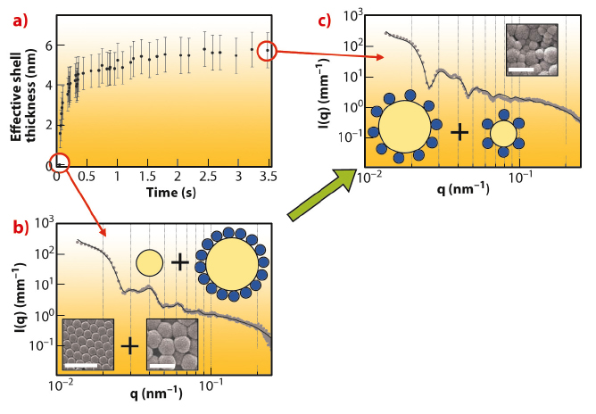
The redistribution of silica nanoparticles that occurs when a P2VP-silica nanocomposite (20 nm silica on a 616 nm sterically-stabilised poly(2-vinylpyridine) latex) is challenged by the addition of a 334 nm sterically-stabilised P2VP latex was also monitored by stopped-flow SAXS (see Figure 57). Here the latex diameters were deliberately chosen to be significantly different because this facilitates the data analysis (Figures 57b and 57c). Silica redistribution is essentially complete within a few seconds after mixing the nanocomposite particles with the sterically-stabilised latex (Figure 57a). There is a rapid initial increase in the effective shell thickness, followed by a period of slower growth until equilibrium is attained after around 2.0 s. The effective thickness of the growing shell was obtained by fitting the SAXS patterns using a three-population model (the additional third population accounts for the second population of smaller core-shell particles).
The experimental time-scales for the adsorption and redistribution of silica nanoparticles compare well with that estimated based on the Smoluchowski fast coagulation rate equation. This suggests that silica exchange is mediated by Brownian collisions. This seems reasonable because the silica nanoparticles are only weakly adsorbed via the steric stabiliser chains, rather than in direct contact with the latex surface (thus avoiding the primary minimum of the potential energy interaction curve).
Our observations are expected to have important implications for the development of nanocomposite paints. Clearly, the performance of such coatings may be compromised if the silica nanoparticles can readily desorb from the latex binder in situ and re-adsorb onto the pigment and/or extender particles present in all modern paint formulations.
Principal publication and authors
J.A. Balmer (a), O.O. Mykhaylyk (a), S.P. Armes (a), J.P.A. Fairclough (a), A.J. Ryan (a), J. Gummel (b), M.W. Murray (c), K.A. Murray (c) and N.S.J. Williams (c), J. Am. Chem. Soc. 133, 826-837 (2011).
(a) Department of Chemistry, University of Sheffield (U.K.)
(b) ESRF
(c) AkzoNobel, Wexham Road, Slough, Berkshire (U.K.)
References
[1] J.A. Balmer, S.P. Armes, P.W. Fowler, T. Tarnai, Z. Gaspar, K.A. Murray and N.S.J. Williams, Langmuir 25, 5339 (2009).
[2] J.A. Balmer, O.O. Mykhaylyk, J.P.A. Fairclough, A.J. Ryan, S.P. Armes, M.W. Murray, K.A. Murray and N.S.J. Williams, J. Am. Chem. Soc. 132, 2166 (2010).
[3] J.A. Balmer, O.O. Mykhaylyk, A. Schmid, S.P. Armes, J.P.A. Fairclough and A.J. Ryan, Langmuir 27, 8075 (2011).


