- Home
- News
- Spotlight on Science
- Exploring the pores...
Exploring the pores in a heavy metal: the 3D microstructure of coatings for nuclear fusion reactors is revealed by high-resolution synchrotron microtomography
06-08-2010
Three-dimensional images of the porous microstructure in plasma-sprayed tungsten are needed to assess whether the material is suitable as a coating for nuclear fusion reactors. High-resolution synchrotron-radiation microtomography (SRµCT) is the only method capable of providing this 3D information - and still the measurements were a challenge.
Share
Vacuum plasma-sprayed tungsten (VPS-W) is a complex-shape porous material. It is a candidate to cover first wall components in future nuclear-fusion reactors [1]. In VPS-W, the refractory nature and low erosion rate of tungsten are combined with a random porous microstructure that strongly affects the thermomechanical behaviour of the material. Accurate micromechanical modelling is required to understand the complex mechanisms governing heat transfer, damage and failure of the coating. Since pore shape and distribution are both highly irregular, their exact influence on the micromechanical properties cannot be modelled analytically. Modelling the structure is therefore a challenging computational task. So far calculations were based on two-dimensional section images obtained by scanning-electron microscopy (SEM). This data was used to generate two-dimensional finite element models [2]. The lack of the third dimension, however, can affect the accuracy of the results, since local stress or the local heat-flux distribution across the 3D solid microstructure can differ from the 2D case. Furthermore, the extraction of three-dimensional parameters from two-dimensional images can be subject to systematic errors.
To improve the reliability of micromechanical calculations, it is therefore necessary to know the 3D microstructure of VPS-W in detail. Since in the real application the coating is always joined to a substrate, the three phase transition zone (steel, tungsten, pores) between coating and substrate is also of great interest.
The aim of the present study was to obtain correct and accurate three-dimensional models from high-resolution synchrotron microtomography. The use of synchrotron radiation was essential here because of the high spatial resolution needed, and because of the possibility to use a monochromatic beam, which facilitates quantitative morphometric analysis because it eliminates the majority of beam hardening artifacts in the data.
 |
|
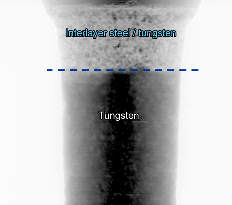
Figure 1. Projection radiograph of a VPS-W sample. On the logarithmic grey scale, dark areas correspond to strong absorption. The dashed line indicates the transition zone between the porous tungsten and the interlayer zone, consisting of steel with tungsten precipitates. |
The experiments were carried out at the ESRF tomography beamline ID19 [3], at a photon energy of 52 keV and with an effective detector pixel size of 1.4 µm. The experiment was challenging because of the high spatial resolution required, in combination with the strong absorption and strong scattering of the X-rays by the material under investigation. Strong absorption can usually be countered by using higher X-ray photon energies; but the attainable spatial resolution degrades with increasing energy. The strong scattering, caused by the porosity of the material on mesoscopic length scales, induces a background signal in the reconstructed volume data. Advanced segmentation algorithms are therefore needed to binarise the volume images, i.e., to assign to each voxel in the volume a label “material” or “void”. The best trade-off between spatial resolution, photon energy and sample size is reached when the cylindrical samples have a diameter of approximately 0.5 mm or less. Unfortunately VPS-W is very brittle, which made sample preparation another challenge and reduced the success rate in sample production to only 15%. Nonetheless, from an original VPS-W coating (PLANSEE A.G., Austria) more than 10 samples were successfully cut by our commercial partner Stangl & Co. (Germany).
Figure 1 shows an X-ray radiograph of one of the samples. Many radiographs of this kind, taken at different viewing angles of the sample, allow tomographic reconstruction of the full volume, as shown in Figure 2, which represents three-dimensional renderings of a segmented region of interest in one of the samples investigated.
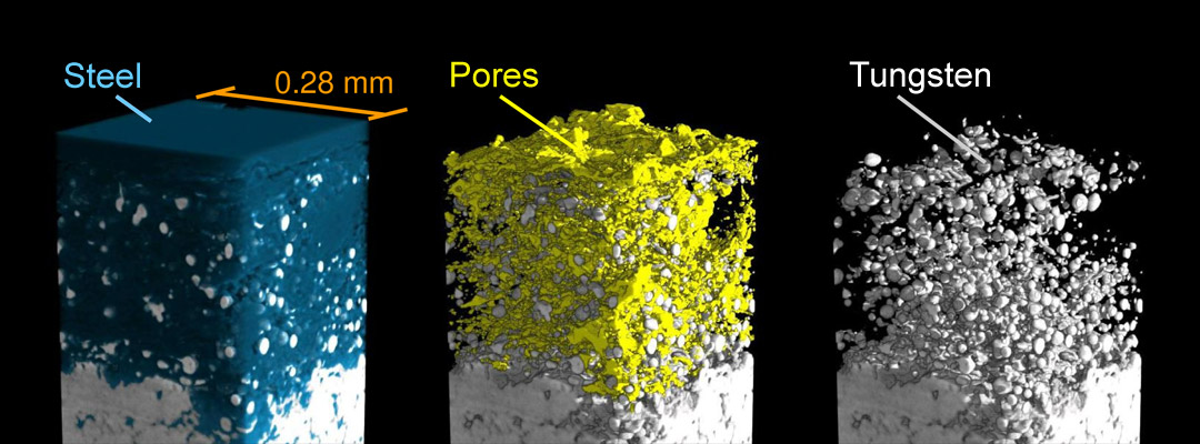 |
|
Figure 2. Three-dimensional renderings of a region of interest from a tomographic volume data set of plasma-sprayed tungsten, obtained at ID19 using 52-keV X-rays (voxel size 1.4 µm). The pictures show the tungsten–steel interlayer. The tungsten layer (light grey) is well attached to the steel substrate (blue); the mixing zone shows an extensive pore network (yellow). |
The results provide genuine high-quality data of the porous microstructure. They can now be used to develop improved thermomechanical finite element models of plasma-sprayed tungsten and thus contribute to the further development of porous tungsten-based materials in general and their application in nuclear fusion technology in particular.
References
[1] H. Greuner, H. Bolt, B. Böswirth, S. Lindig, W. Kühnlein, T. Huber, K. Sato and S. Suzuki, Fusion Engineering and Design 75-79, 333-338 (2005).
[2] A. Zivelonghi et al., J. Nucl. Mater., accepted.
[3] T. Weitkamp, P. Tafforeau, E. Boller, P. Cloetens, J.-P. Valade, P. Bernard, F. Peyrin, W. Ludwig, L. Helfen and J. Baruchel, AIP Conf. Proc. 1221, 33-38 (2010).
Authors
A. Zivelonghi (a), T. Weitkamp (b), J.-H. You (a), C. Linsmeier (a), M. Rasinski (a), G. Weizmann (a), M. Schlüter (a), C. Capuano (c).
(a) Max-Planck-Institut für Plasmaphysik, Garching (Germany)
(b) ESRF
(c) ITER
Top image: The 3D microstructure of a vacuum plasma-sprayed tungsten coating system. Composite image to show the distribution of the pores within the tungsten (bottom) and tungsten/steel interlayer (top).
partners


European Synchrotron Radiation Facility - 71, avenue des Martyrs, CS 40220, 38043 Grenoble Cedex 9, France.