- Home
- News
- Spotlight on Science
- Fusion reactor pebble...
Fusion reactor pebble bed studied by X-ray tomography
24-09-2010
The thermo-mechanical behaviour of pebble beds in the blanket of a fusion reactor depends on pebble arrangement in the beds, the number of pebble-pebble and pebble-blanket wall contacts, as well as the respective contact surfaces. The safe operation of the blanket requires efficient tritium release throughout the gas percolation pathways inside the pebbles. High-resolution microtomography provides valuable 3D information about such a system.
Share
Understanding the thermo-mechanical behaviour of the beryllium pebble beds in the helium cooled pebble bed (HCPB) blanket design is crucial for future fusion reactors such as ITER. Accurate knowledge is needed of the inner structures of the pebbles, since helium and tritium bubbles are produced inside the pebbles during the life cycle of a power reactor. The tritium build up can be as much as several kilograms, therefore becoming a key safety issue. Non-destructive analysis to monitor the arrangement of pebbles as well as the morphology of gas bubbles in neutron irradiated pebbles were carried out on ESRF beamlines ID15, ID17 and ID19, based on 3D computer aided microtomography with high spatial resolution.
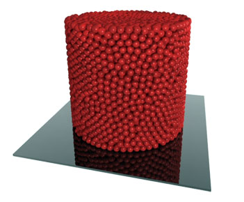
Aluminium cylindrical capsules filled with equally sized spherical aluminium pebbles (used instead of Be pebbles for better contrast) of 2.3 and 5 mm diameter, like the one depicted in Figure 1, were used as samples for the tomography measurements [1]. The experiments were performed with a pink beam centred around 80 keV and an effective detector pixel size of 14.4 µm.
 |
|
Figure 1. Photorealistic 3D rendition of the reconstructed volume of a pebble bed sample. Sample inner diameter D=49 mm, height H≈46 mm, diameter of the aluminium spheres d=2.3 mm. |
Using the three-dimensional images obtained by tomography, the number of contacts between the pebbles and their angular distribution were determined as well as the related contact surfaces. Some of the pebble capsules were subjected ex situ to high pressure (from 2 to 16 MPa) before the experiment, simulating the effect of pressure build-up in the pebble bed during blanket operation. It could be shown that the pebble contact areas with walls become so large under blanket relevant conditions that an additional heat transfer resistance between pebble bed and wall can be neglected. Moreover, it was found that the axial void fraction profiles for compressed beds differ characteristically from those for non-compressed beds due to frictional effects within the pebble bed. Another important finding is that the layer of the spheres in contact with the bottom plate of the capsule exhibits domains with hexagonal structure in the inner region. At increasing distance from the bottom, the hexagonal structure domains become smaller and eventually vanish where the fourth regular layer should be located, turning into a fairly homogenous distribution of pebble contacts in the bulk.
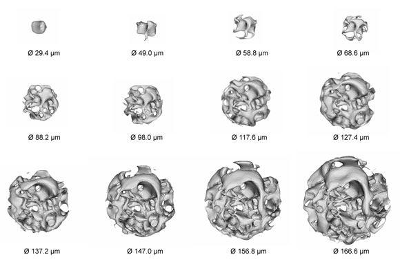
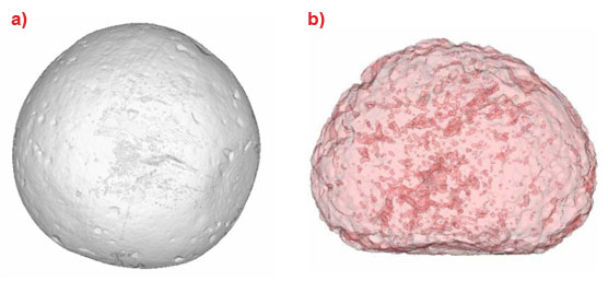
The tomographic scans on individual beryllium pebbles, both freshly fabricated and neutron irradiated, were performed at 4.9 and 1.4 µm resolution using a monochromatic beam of 10 keV [2]. Prior to the tomography measurements, some pebbles had been submitted to a fast neutron fluence of 1.24×1024 m-2 in a nuclear reactor, which corresponds approximately to 480 atomic parts per million (appm) 4He and 12 appm 3H production. After irradiation, the pebbles were annealed at high temperature (1500 K) to vent most of their tritium content. The evaluation of bubble sizes and densities in single beryllium pebbles, the assessment of the pore channel network topology, the 3D reconstruction of the fraction of interconnected bubble porosity, and the open-to-closed porosity ratio are among the most interesting findings of this investigation. In the fresh pebbles the size of fabrication-induced voids was found to range between 9 and 18 µm, causing a total void fraction no higher than 0.17%. In the irradiated and annealed samples, besides a bimodal pore distribution with a smaller population of about 10 µm diameter and a high density of almost entirely (99%) interconnected pores around 40 µm diameter, a swelling of 14% was found (see Figure 2). The typical distances between percolation paths as well as their pattern suggest that they have been formed primarily along the grain interfaces. The complexity of the porosity network in an irradiated pebble is apparent from Figure 3, where its interior is explored by means of regions of interest which are successively inscribed concentric spheres, starting from a minimum diameter of 29.4 µm up to a maximum diameter of 166.6 µm [3]. It should be noted that only the porosity topology is represented by the grey-scale.
 |
|
Figure 3. The void spatial distribution complexity in a beryllium pebble highlighted by successively inscribed spherical ROIs of increasing size from a diameter of 29.4 µm up to 166.6 µm. |
X-ray microtomography provided an essential aid not only for the experimental validation of the multiscale modelling of gas release kinetics but also for the assessment of the structural integrity of entire pebble bed assemblies.
References
[1] J. Reimann, R.A. Pieritz, C. Ferrero, M. di Michiel, R. Rolli, Fusion Engineering and Design 83, 1326–1330 (2008).
[2] A. Möslang, R.A. Pieritz, E. Boller, C. Ferrero, Journal of Nuclear Materials 386–388, 1052–1055 (2009).
[3] R.A. Pieritz, J. Reimann, C. Ferrero, Advanced Engineering Materials, accepted.
Authors
J. Reimann (a), R.A. Pieritz (b), C. Ferrero (c), M. di Michiel (c), A. Möslang (a), P. Tafforeau (c), E. Boller (c)
(a) Karlsruher Institut für Technologie, Karlsruhe (Germany)
(b) ARS - Applied Research Solutions, Chirens (France)
(c) ESRF
Top image: Tomography shows the pores within a beryllium pebble


