- Home
- News
- Spotlight on Science
- Mapping disorder...
Mapping disorder with X-rays
07-03-2011
The structure of disordered surfaces at the atomic level has remained elusive. Now, using a combination of inelastic X-ray scattering in grazing-angle geometry and simulations, a team of European researchers has gained – for the first time – insight into the arrangement of chemical bonds at the surface of a simple, mono-atomic glass: amorphous selenium.
Share
Investigation of the crystalline structure and atomic order at surfaces by microscopic and scattering techniques reveals valuable information for fields ranging from nanotechnology to catalysis. The periodic arrangement of their constituents permits such ordered surfaces to be imaged in amazing detail. Diffraction methods, which exploit the reciprocal space rather than real space, typically give excellent spatial resolution. However, this is not the case for disordered surfaces where the missing translational symmetry of the surface prohibits the definition of a finite unit cell and limits available techniques to direct imaging methods. Although direct imaging methods such as atomic force microscopy should give comparable resolution for both ordered and disordered surfaces, practice shows that atomic contrast is feasible only on crystalline surfaces.
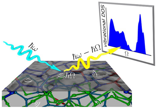
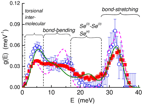
A team of researchers from Italy, Austria, Greece, France and the Czech Republic has taken a new approach to the investigation of the surface structure of disordered materials. In the first step they performed inelastic X-ray scattering experiments at grazing incidence at beamline ID28 in order to discover the vibrational spectrum of the Se-glass surface. To be surface-sensitive, the incidence angle was below the critical angle of total external reflection (Figure 1). In the second step, they analysed the vibrational features in the spectrum and interpreted them using ab initio and semi-classical simulation approaches (Figure 2).
The researchers found that Se atoms frequently arrange in pairs of 3-fold and 4-fold bonded atoms in the surface layer, which is in apparent contrast to the bulk where Se atoms preferentially make bonds with two neighbours only, thus forming long one-dimensional chains. This so-called branching of the one-dimensional chains in a rather extended surface layer of 5 nm or more is responsible for the formation of a “protective” layer with enhanced rigidity. In turn, the higher rigidity of the surface layer is expected to affect the functionality and responsiveness of the surface.
Principal publication and authors
T. Scopigno (a,b), W. Steurer (c), S.N. Yannopoulos (d), A. Chrissanthopoulos (d,e), M. Krisch (f), G. Ruocco (a), and T. Wagner (g), Vibrational dynamics and surface structure of amorphous materials, Nature Communications 2, 195 (2011).
(a) Dipartimento di Fisica, Universitá di Roma 'La Sapienza' (Italy)
(b) IPCF-CNR, UOS Roma, c/o Dipartimento di Fisica, Universitá 'Sapienza', Roma (Italy)
(c) Institute of Physics, Surface and Interface Physics, Karl-Franzens University, Graz (Austria)
(d) Foundation for Research and Technology—Hellas, Institute of Chemical Engineering and High-Temperature Chemical Processes (FORTH/ICE–HT), Patras (Greece)
(e) Department of Chemistry, University of Patras (Greece)
(f) ESRF
(g) Department of Inorganic Chemistry, Faculty of Chemical-Technology, University of Pardubice (Czech Republic)
Top image: Representation of the outermost skin of a-Se surface from a fully-optimised 96-atom Se structural model. Colour of Se atom indicates branching, red: four-fold; blue: 3-fold; light green, two-fold; dark green: Se atom terminated by H-bond; grey: H atom.


