- Home
- News
- Spotlight on Science
- Observing a target...
Observing a target enzyme of Alzheimer’s disease drugs in action
03-11-2008
Acetylcholinesterase is one of the main targets for drugs treating Alzheimer’s disease. Scientists at the Institut de Biologie Structurale in Grenoble have carried out experiments at the ESRF to observe acetylcholinesterase in action with the aim to understand its molecular mode of action. They observed structural changes in one of Nature’s fastest enzymes during its catalytic cycle. Understanding the structural dynamics of acetylcholinesterase is an important prerequisite for the design of more efficient drugs.
Share
The enzyme acetylcholinesterase plays an essential role in the process of nerve signal transmission at cholinergic synapses where it rapidly breaks down the neurotransmitter acetylcholine. In Alzheimer’s disease, loss of nerve signal transmission results in the impairment of mental capacity (memory, judgement, language, etc.) that is associated with the disease. Consequently, many treatments for Alzheimer’s disease involve drugs targeting acetylcholinesterase. The drugs are inhibitors of acetylcholinesterase and permit greater concentrations of acetylcholine at nerve junctions.
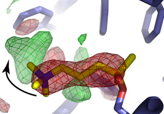
Researchers at the Institut de Biologie Structurale in Grenoble, the Institut de Pharmacologie et de Biologie Structurale in Tolouse, the Weizmann Institute of Science in Israel, and the ESRF were able to observe acetylcholinesterase in the process of cleaving the neurotransmitter. They used a novel method that allowed both initiation and monitoring of the reaction by the intense X-ray beam provided by the ESRF. To observe the enzyme at work, an analogue of acetylcholine within the enzyme active site was cleaved by the X-ray beam in a fashion that simulates enzymatic catalysis of the neurotransmitter by acetylcholinesterase. The latter could then be observed by X-ray crystallography in a conformation that it would adopt during catalysis. However, catalysis by acetylcholinesterase normally occurs in less than one thousandth of a second at room temperature. Hence the experiments were conducted at temperatures between -120 and -170°C. This was key to slowing down the reaction so that two successive enzymatic conformations could be observed (Figure 1).
 |
|
Figure 1. After cleavage of an analogue of the neurotransmitter acetylcholine (yellow sticks) in the active site of acetylcholinesterase (blue sticks), choline, one of the cleavage products, reorients in the enzymatic active site. This reorientation, from the red to the green position, is indicated by the black arrow. |
The strategy used to trigger and monitor the reaction is of particular interest as it made use of specific radiation damage during a temperature controlled crystallographic experiment. This method could be applied to other systems where a suitable substrate analogue is available.
Certain drugs act by blocking their target in a specific conformation. Knowing the ensemble of conformations accessed by a macromolecular target can thus aid in the rational design of more potent drugs. The results open up novel approaches to the design of second-generation drugs to treat Alzheimer's disease.
Principal publication and authors
J.-P. Colletier (a, b), D. Bourgeois (a, c), B. Sanson (a), D. Fournier (d), J.L. Sussman (e), I. Silman (e) & M. Weik (a), Shoot-and-trap: use of specific X-ray damage to study structural protein dynamics by temperature-controlled cryo-crystallography, Proc. Natl. Acad. Sci. 105, 11742 (2008).
(a) Institut de Biologie Structurale, Grenoble (France)
(b) University of California, Los Angeles (USA)
(c) European Synchrotron Radiation Facility, Grenoble (France)
(d) Institut de Pharmacologie et de Biologie Structurale, Toulouse (France)
(e) Weizmann Institute of Science, Rehovot (Israel)


