- Home
- Industry
- Industry news
- Research centre...
Research centre for the application of steel dives into the dark-field microscopy at ESRF
11-05-2017
Dark-field microscopy offers new perspectives for research in metallurgy. The proof is in the pudding: a new collaboration between OCAS, a Belgian research centre active in metallurgy, and the ESRF, is starting to bear fruits.
Share
Sometimes great alliances come from fortuitous encounters. Back in June 2015, the ESRF organised a workshop on “Non-conventional imaging” for industry. Roger Hubert, Chief Scientific Officer at OCAS, joined in. He had never used a synchrotron before and came across ESRF scientist Carsten Detlefs. They discussed the benefits of using hard X-rays in metallurgical research.
Fast forward 18 months and Can Yildirim, a post-doctoral researcher funded by OCAS, is doing his first experiment on ID06. Hubert explains the benefits of synchrotron radiation in their studies: “The idea to get insights of the inside is very exciting! In most metallurgical studies we look at final microstructures in 2D. When I heard that we could envisage following metallurgical reactions live and in 3D, I was immediately convinced that this was the way to go.”
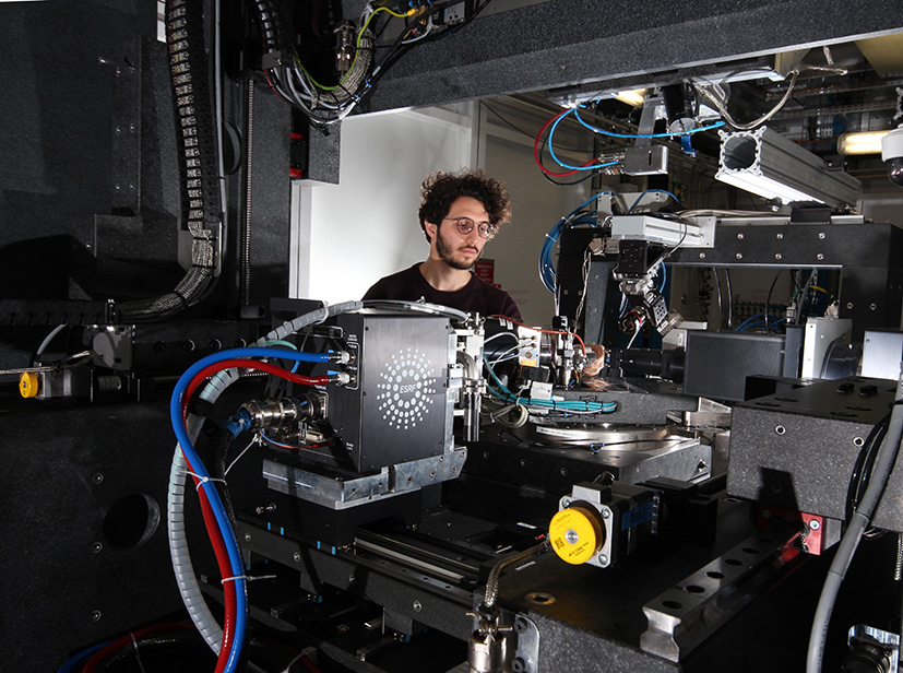
At the ESRF, Yildirim is using the dark-field X-ray microscopy technique for the first time on iron nitrides precipitated in steel. These model samples are used to design the methodology, and especially, the heat treatment cycle management in alloys. In conventional steel processing, nitrogen alloying is challenging due to its limited solubility during casting and solidification. Alternatively, nitriding as thermochemical treatment on the final material (also called case hardening) can be used to significantly improve surface and bulk properties. Controlling the nucleation and growth of nitride phases is essential for macroscopic properties of these alloys. Dark-field X-ray microscopy, through objective lenses, converts diffracted signals to real space images. “We are basically able to see the internal structure of steel alloys and follow which processes occur on sub-micron scale in operando conditions”, explains Can Yildirim.
Apart from this project on iron nitrides, OCAS is also envisaging researching different types of steel alloys, including steels used in transformers and electrical motors.
Come to the Dark Side. We have diffraction!Unlike bright field microscopy, where the sample is seen on a bright background, dark field microscopy detects only light that is scattered by the sample, not from direct illumination. In case of dark-field x-ray microscopy, the signal originates from Bragg diffraction. This new technique is therefore ideal for crystalline materials like most metals, ceramics, rocks, ice, semiconductors, etc. Many physical and mechanical properties of these materials depend on their internal structure, organised into grains of different sizes. Dark-field X-ray microscopy is a non-destructive technique which allows 3D mapping of orientations and stresses from 100 nanometres to 1 millimetre. The technique allows zooming in and out in both direct and angular space. In the case of alloys, scientists can now monitor what happens inside a sample as it undergoes thermal processes. Carsten Detlefs, beamline responsible of ID06 explains that “Dark field x-ray microscopy is inspired by dark-field transmission electron microscopy (TEM). It combines elements from x-ray diffraction topography, x-ray tomography and x-ray microscopy. Compared to TEM, we have a much better angular resolution, although a not as good spatial resolution. What is more important, however, is that we can look inside the material, whether it is inside a furnace or other sample environment, and see how the sample evolves during processing. That is what makes it ideal for industrial clients like OCAS”. With the Extremely Brilliant Source upgrade coming up, the technique will become even more powerful. “We have proposed this as one of the Upgrade Beamlines and hope to open a general user programme about 4 or 5 years from now”, says Detlefs. Find out more about this technique here: Simons, et al, MRS Bulletin, 2016. |
 |
|
Can Yildirim on the ID06 beamline. Credits: Jeff Wade. |
Top image: Alloys go through different thermo-mechanical processes to adjust their properties. Credits: OCAS.


