- Home
- Users & Science
- Scientific Documentation
- ESRF Highlights
- ESRF Highlights 2000
- Chemistry
- The Structural Complexity of a Polar, Molecular Material Brought to Light by Synchrotron Radiation
The Structural Complexity of a Polar, Molecular Material Brought to Light by Synchrotron Radiation
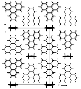
Crystalline materials are often built less regularly than the pictures in most textbooks would suggest. Different unit cells differ in molecular occupation, orientation or position. In spite of such disorder, or sometimes because of it, such materials may have interesting properties. Molecular inclusion compounds of the hydrocarbon host molecule perhydrotriphenylene (PHTP) with rod-shaped, polar guest molecules, e.g. nitrophenylpiperazine (NPP), provide an example [1]. Stacks of host molecules are arranged in a honeycomb architecture whose tunnels are filled with chains of guest molecules (Figure 16). Right- and left-handed host molecules occupy the stacks in a not-quite-random fashion. The chains of guest molecules register at different heights in different channels. Remarkably, they are all parallel, even though the channels are ~ 15 Å apart. The crystals are polar and show second harmonic generation.
 |
Fig. 16: The structure of PHTP 5.NPP viewed down the polar stacking axis of PHTP host and NPP guest molecules; z = 0.75 for shadowed PHTP molecules, z = 0.25 otherwise; z = 0, 1, 2, 3 or 4 for NPP molecules. The wavy shapes of right- and left-handed, D 3-symmetric PHTP molecules are indicated at the right margin of the unit cell. The long axis of NPP molecules is perpendicular to the plane of projection.
|
This material produces an extraordinarily rich diffraction pattern including Bragg peaks and incommensurate satellites, as well as diffuse streaks, planes and three-dimensional features. Almost complete patterns have been measured on beamline BM1A (SNBL) at the centre and at one tip of a prismatic crystal (298 K and 120 K). Five gigabytes of accurate, raw data obtained with an area detector in 16-bunch mode have been processed into 400 layers in reciprocal space [2].
Most of the diffuse and satellite scattering can be assigned to occupational disorder of the host, positional disorder of the guest, or to local distortions of the average structure. Real structures are simulated with a Markov growth model. Its parameters are optimised with an evolutionary algorithm on 40 individuals using an R-value based on diffraction intensities as fitness criterion. The structure simulations and Fourier transformations are calculated through a scheme of distributed computing with up to ten simultaneous processes running on PC's, SUN and SGI workstations and progressing at a rate of ~ 15 generations per day.
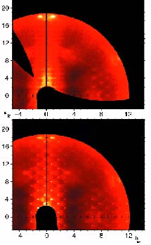
Figure 17 shows a particularly interesting feature. It compares the scattering of 100 µ slabs in the centre and at a tip of the prismatic crystal. In the centre the diffraction symmetry is nearly orthorhombic (top), whereas at the tip it is monoclinic (bottom). This implies that the crystal is not homogeneous along the stacking direction of the host and guest molecules. Note that at the crystal tip the satellite reflections are connected through triangular diffuse scattering. The former indicate a well defined long-range ordered, incommensurate modulation of the structure, the latter some dispersion of this modulation.
 |
Fig. 17: hk2.4 layer from the centre (top) and a tip (bottom) of a PHTP 5.NPP crystal at 120 K. Note that diffuse intensities form a pseudo-rectangular pattern at the crystal centre, but are triangularly shaped at its tip.
|
Two conclusions emerge: (1) accurate and complete three-dimensional diffuse scattering data of a complex disordered molecular material can be measured with synchrotron radiation and three-dimensional structure models developed with a scheme of distributed computing. The scope of such studies becomes significantly broader if the computations are parallelised. (2) Diffuse scattering and thus disorder of the crystal structure along growth directions may be followed. With an improved focusing device, spatial resolution could be significantly improved, thus providing records of the crystallisation history of disordered materials.
References
[1] O. König, H.B. Bürgi, Th. Armbruster, J. Hulliger and Th. Weber. J. Am. Chem. Soc., 119, 10632 (1997).
[2] M.A. Estermann and W. Steurer. Phase Trans., 67, 165 (1998).
Principal Publication and Authors
T. Weber (a), M.A. Estermann (b), H.B. Bürgi (a), Acta Cryst. Sect. B, accepted.
(a) Universität Bern (Switzerland)
(b) ETHZ, Zürich (Switzerland)


