- Home
- Users & Science
- Scientific Documentation
- ESRF Highlights
- ESRF Highlights 2004
- X-ray Imaging
- X-ray Microtomographic Study of Liquid Foams
X-ray Microtomographic Study of Liquid Foams
Fluid foams consist of gas bubbles separated by a continuous film of liquid which tends to minimise its surface energy. They are a model for various classes of materials: emulsions, magnetic garnets, and even grain boundaries in crystals [1]. The knowledge we gain from understanding foams has technological applications, ranging from food and shaving cream to fire fighting and oil recovery. Understanding the structure is an important step towards predicting a foam's mechanical properties. Moreover, the structure of fluid foams determines the structure of solid foams produced by solidification: metallic foams for the automobile industry, pumice from volcanoes, polymeric foams as filling materials and food foams.
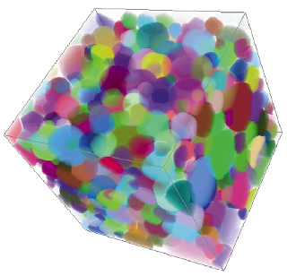
X-ray microtomography is by far the best non-invasive method to look inside a medium which scatters visible light as much as foams do. Since X-rays pass through the foam, a raw picture is a 2D projection of the real 3D structure. The idea of tomography is to take typically 1000 pictures of the same sample at different angles. A mathematical inversion yields the 3D reconstruction of the whole foam. However, as a liquid foam can evolve, it is important to take the 1000 pictures in a short time to avoid blurring in the reconstruction. The ESRF's brightness and beamline improvements on ID19 have enabled us to reduce the acquisition time from hours (which was fine for solid structures) to tens of minutes, then to minutes, with a resolution down to 10 or even 1 µm (Figure 141).
 |
Fig. 141: Reconstructed 3D image of the 1 cm3 liquid foam (air bubbles in soap solution): a billion of 10-µm voxels (volume image elements). The (false) colours label bubbles. |
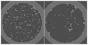
We can obtain a wealth of details. On the smallest scale, we can observe thin water films, including the accumulation of water in the borders where films meet. On the scale of bubbles, we can quantify their size, shape, and number of neighbours. Since we track thousands of bubbles individually, we can perform statistics on the scale of the whole foam, and even obtain correlations, for instance between size and number of neighbours. Finally, we can observe a phenomenon common to emulsion and grain growth in metals: since the air can slowly diffuse through water, it flows from bubbles at high pressure (actually, the smallest ones) to low pressure regions (large bubbles). Thus small bubbles shrink, and eventually disappear. The total number of bubbles decreases, and in time (few hours) the average length scale coarsens (Figure 142) [2]. Preliminary tests with a new Dalsa camera yielded images of an even dryer foam (that is, with more air and thinner walls) in a few tens of seconds; this opens the way to the dynamic study of evolving foams.
 |
Fig. 142: 2D section of the foam after ageing 1 hour (left) and 1 day (right). |
References
[1] D. Weaire, S. Hutzler, Physics of Foams, Oxford Univ. Press, 1999.
[2] Movies available at http://www-lsp.ujf-grenoble.fr/link/mousses-films.htm
Principal Publication and Authors
J. Lambert (a), I. Cantat (a), R. Delannay (a), A. Renault (a), F. Graner (b), J.A. Glazier (c), I. Veretennikov (c), P. Cloetens (d), Colloids Surf. A: Physicochem. Eng. Aspects, in press.
(a) GMCM, Univ. Rennes 1 (France)
(b) LSP, Univ. Grenoble 1 (France)
(c) Dept Physics, Indiana Univ. (USA)
(d) ESRF


