- Home
- Users & Science
- Scientific Documentation
- ESRF Highlights
- ESRF Highlights 2004
- Soft Condensed Matter
- GID Studies of Oriented Nucleation of Semiconductor Nanoparticles on Polyconjugated Langmuir Films at the Air-Solution Interface
GID Studies of Oriented Nucleation of Semiconductor Nanoparticles on Polyconjugated Langmuir Films at the Air-Solution Interface
Polyconjugated Langmuir films of polydiacetylene (PDA) are anisotropic, robust films that change their colour from blue to red upon thermal, mechanical, UV and shorter wavelength irradiation or chemical stimulation. Parallel polymer backbones are organised in 2-D crystalline domains whose typical size varies from several µm to several hundreds of µm. In previous work, we used TEM and AFM to evaluate the lateral ordering and crystallographic orientation of nanocrystalline PbS, CdS and Ag2S deposited on PDA LF templates [1-4]. These studies involved transfer of the ultrathin films onto solid supports and their study ex situ. This may involve structural changes during the transfer. Evidently, in situ grazing-incidence X-ray diffraction (GID) structural studies at the air-solution interface can overcome this problem, elucidate the true structure of the films and allow the study of the factors affecting it. 10,12-pentacosadiynoic acid in chloroform solution was spread over an aqueous subphase in a Langmuir trough mounted at beamline ID10B. After the solvent has evaporated, the monomers were compressed to their stable trilayer structure and polymerised by ultraviolet irradiation.
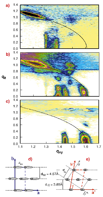
We monitored the transition from the blue to the red forms of the PDA Langmuir films. Accurate control of radiation (combined synchrotron and UV) dose was achieved by lateral translation of the trough with respect to the beam during scans. Hence, any given area in the sample can be varied from very short to longer exposure time periods. GID maps (reflected intensity, vs qxy and qz) of the pure blue and red phases and their mixtures were obtained (Figure 65a-c). The pure blue phase, obtained by minimal exposure, is shown in Figure 65a. Continued exposure to X-ray radiation gradually increases the relative amount of the 'red' phase (Figure 65b). These GID maps depict coexistence of the two phases. Upon further increase in radiation dose, the entire film converts to the 'red' phase (Figure 65c). The transition between the phases appears to be discrete, with no intermediate structures observed. The 'blue' to 'red' conversion is manifested in a shift of the qxy and qz position of the two Bragg peaks obtained from the centred rectangular structure to higher qxy values. Note also the shift in position of the intense reflection at qxy = 1.18 and qz = 1.1 [Å-1] along the same qtotal arc to circa qxy = 1.60 [Å-1] and qz close to zero (near-normal orientation of the alkyl side groups) from Figure 65a to Figure 65c. Based on these results, we constructed a model describing the crystallographic unit cells corresponding to each phase, as shown in Figure 65d,e.
 |
Fig. 65: GID maps depicting the gradual structural transformation of PDA blue phase (a), through coexistence situation (b) to a fully transformed 'red' phase (c). The black arc corresponds to qtotal = 1.6 [Å-1]. The reconstructed unit cell of the blue phase (d) and the red phase (e) are shown. The red phase is denser with the alkyl chains nearly vertical. |
PDA-PbS nanocrystal hybrid materials were produced by exchanging the pure water subphase with a solution containing Pb2+ cations and subsequently exposing the interface to controlled amounts of H2S gas. This resulted in the formation of triangular and elongated PbS nanocrystals, aligned in linear rows along the polymer strands as shown in the TEM image in Figure 66a, inset. The equidimensional triangular crystallites are (111) oriented. The right angle and the elongated crystals are (100) oriented. Both crystal orientations are aligned with the [110] direction along the PDA backbone[4]. In situ GID studies were carried out in order to compare the orientation of the PbS nanocrystals in the presence or absence of PDA template films at the interface. GID diffraction pattern from the hybrid material depicts the contribution of the organic template and both (100) and (111) orientations of the incipient crystallites (Figure 66a). In the absence of PDA, only the (100) orientation is observed, as expected for crystals with the rocksalt structure (Figure 66b). The (111) orientation, directly induced by the PDA film, is a result of the strong interactions between the PDA carboxylic headgroups and the metal cations in solution. The GID study of templated crystallites under the film indicated strong association with the red phase of PDA. These metal cation LF interactions are currently being studied in more detail by the authors.
 |
Fig. 66: GID maps of (a) PbS nanocrystals nucleated under PDA film. Reflections from the PDA layer are marked with a frame. Inset: TEM micrograph depicting the PbS on PDA. (b) GID map of PbS grown at the air-solution interface in the absence of PDA, indicating that all crystals are (100) oriented. Inset: PbS crystals in the absence of PDA. |
References
[1] A. Berman, and D. Charych, Adv. Mater 11, 296 (1999).
[2] N. Belman, A. Berman, Y. Lifshitz, V. Ezersky and Y. Golan, Nanotechnology 15, S316 (2004).
[3] A. Berman, N. Belman and Y. Golan, Langmuir 19, 10962 (2003).
[4] N. Belman, Y. Golan and A. Berman, Crystal Growth and Design (2004), in press.
Principal Publication and Authors
A. Berman (a), Y. Lifshitz (a), O. Konovalov (b) and Y. Golan (a).
(a) Ben Gurion University (Israel)
(b) ESRF


