- Home
- Users & Science
- Scientific Documentation
- ESRF Highlights
- ESRF Highlights 2004
- Surface and Interface Science
- Seeing Adsorbed Hydrogen Atoms on Metals with Surface Diffraction: Towards Catalytic Conditions
Seeing Adsorbed Hydrogen Atoms on Metals with Surface Diffraction: Towards Catalytic Conditions
One of the reasons for the quite late appearance of in situ studies of model catalysts "in action" by Surface X-ray Diffraction (SXRD) [1] is the widespread belief that surface diffraction can not "see" light adsorbed atoms and molecules, coming from the gas phase above the catalyst. This belief was already undermined by articles showing that the diffraction signals from an ordered layer of adsorbed CO molecules [2] or from a light oxide surface (the "zero electron" signals, which are proportional to (fMg2+-fO2-), from a MgO(001) surface) [3], could easily be detected.
Here we present further evidence of the sensitivity of SXRD to light atoms, on the textbook case of a p(2x2) ordered overlayer of hydrogen atoms on Ni(111). We also briefly describe the effect, relevant for catalysis, of exposure of the Ni(111) surface to one bar of H2.
 |
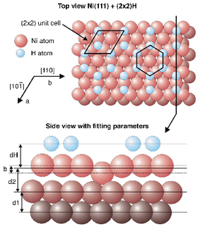
Fig. 86: The model of the UHV low-T p(2x2) structure of H adsorbed on Ni(111) proposed in [4]. The fitting parameters of the atomic model are shown. The values of these parameters found from our SXRD dataset compare well with the ones found from the LEED study [4]. |
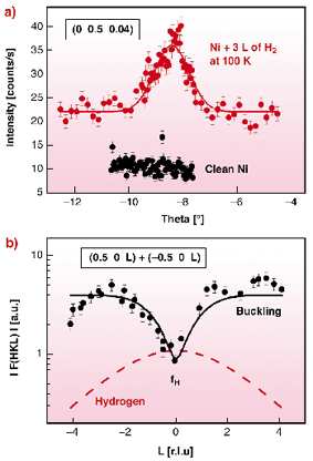
Figure 86 shows the atomic model proposed by Hammer et al. [4] for the Ultra-High Vacuum (UHV) low-temperature p(2x2) structure of H2 on Ni(111), using quantitative Low Energy Electron Diffraction (LEED). This structure consists of half a monolayer of H atoms, with the Ni atoms located below the H atoms moving upwards (buckling). Figure 87 shows a part of the SXRD dataset collected on ID03 for this same structure. This structure is a good test-case of the sensitivity of SXRD to H atoms, because there are locations in reciprocal space where the diffracted intensity is purely due to H scattering. The low-L part of the fractional order rods (Figure 87a) is one such location, for two reasons. First the buckling (z-motion of the Ni atoms) of the last Ni plane contributes only at non-zero L's (Figure 87b), and secondly, the symmetry of the model (Figure 86) forbids x-y motions of the Ni atoms. The intensity due to H scattering in Figure 87a is 20 cts/s above background, with a full-width of 1. The fitting of the structural parameters shown in Figure 86, using the SXRD data, gives values that agree well with the ones of [4]. The biggest error bar, as in the LEED study, is on the z-position of the H atoms, due to the very small L-range of proper interference between Ni and H scattering.
 |
Fig. 87: (a) rocking scan around one of the SXRD peaks from the p(2x2) H-layer at 100 K. The scan after adsorbing H (top) is compared to the scan on the clean Ni (bottom). At this low L value, the peak comes purely from scattering by H atoms. (b) One of the fractional order reconstruction rods. The data (points) are compared to the calculated rod (solid line). The contribution of H atoms is shown by the dashed line. It is the main contribution at low L. |
Room temperature exposure to H2 at pressures ranging from 10-8 mbars to 1.1 bar was also studied. In this case, the surface stays (1x1) upon adsorption, i.e. there is no ordered superstructure of the H atoms. The surface structure remains practically the same as under UHV up to 300 mbar. Above 300 mbars, a slight decrease (max -40%) of the Ni crystal truncation rods occurs, interpreted by a mixture of surface disorder and/or roughness, possibly due to H embedding. This is an important result as it predicts a possible change at 300 mbars in the catalytic properties of Ni for the methanation reaction.
References
[1] M.D. Ackermann, O. Robach, C. Walker, C. Quirós, H. Isérn, S. Ferrer, Surf. Sci. 557, 21 (2004).
[2] I.K. Robinson, S. Ferrer, X. Torrelles, J. Alvarez, R. Van Silfhout, R. Schuster, R. Kuhnke, K. Kern, Europhys. Lett. 32, 37 (1995); C. Quirós, O. Robach, H. Isérn, P. Ordejon, S. Ferrer, Surf. Sci. 522, 161 (2003).
[3] O. Robach, G. Renaud, A. Barbier, Surf. Sci. 401, 227 (1998).
[4] L. Hammer, H. Landskron, W. Nichtl-Pecher, A. Fricke, K. Hein, K. Müller, Phys. Rev. B 47, 15969 (1993).
Principal Publication and Authors
S. Ferrer (a,b), C.J. Walker (a), M. Ackermann (a,c), C. Quirós (a,d), H. Isérn (a), O. Robach (e), submitted to Phys. Rev. B.
(a) ESRF
(b) ALBA - Edifici Cíencies, Bellaterra (Spain)
(c) Kamerlingh Onnes Laboratory, Leiden University (The Netherlands)
(d) Depto. de Física, Faculdad de Ciencias, Universidad de Oviedo (Spain)
(e) CEA-Grenoble, DRFMC/SI3M/PCM (France)


