- Home
- Users & Science
- Scientific Documentation
- ESRF Highlights
- ESRF Highlights 2007
- Structural Biology
- The crystal structure of bacteriophage T7 endonuclease I resolving a Holliday junction
The crystal structure of bacteriophage T7 endonuclease I resolving a Holliday junction
The rearrangement, or recombination, of DNA is an ancient and ubiquitous biological process. Recombination is central to many biological processes such as the generation of genetic variation (and therefore evolution) and the incorporation of viral DNA into host DNA, resulting in successful infection.
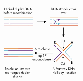
The process of DNA recombination occurs in distinct stages (Figure 78) with the formation of a four-way (Holliday) junction being a central step. For the reaction to proceed the Holliday junction must be cleaved to yield 2 linear duplexes. This process is regulated by a junction-resolving enzyme that cleaves the Holliday junction resulting in rearranged DNA strands. Bacteriophage T7 encodes a protein, endonuclease I, which has been shown to act as a Holliday junction resolving enzyme.
 |
|
Fig. 78: The process of recombination. |
To understand the mechanism by which endonuclease I binds and cleaves Holliday junctions, we have solved the structure of a complex of endonuclease I and a synthetic 4-way DNA junction using X-ray crystallography. To obtain high quality X-ray diffraction data we had to purify 17 different Holliday junction complexes, set up 25,000 crystallisation trials and collect data from approximately 200 crystals at beamline ID23-2. Our best data extended to 3.1 Å resolution and the structure of the complex was solved by molecular replacement using the structure of DNA-free endonuclease I [1] as the search model.
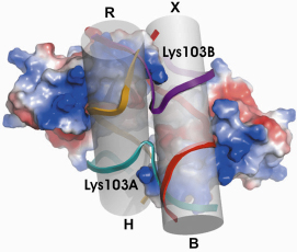
In the complex (Figure 79), pairs of DNA helical arms are essentially stacked on top of one another (R stacked on H and X stacked on B) to form two pseudo-continuous duplexes with an angle between them of -80°. This represents an alteration of ~ 130°, from the right-handed stacked X-structure of the free Holliday junction in solution (interaxial angle ~ +50°) [2] and is consistent with studies of the structure of the endonuclease I - Holliday junction complex in solution. The observed conformation creates an almost continuous deep cleft across the major groove face of the junction which forms the principal binding site for the protein.
Significant induced fit occurs in the complex, with changes in the structure of both the protein and the junction. The enzyme is a dimer of identical subunits and presents two equivalent binding channels that contact the backbones of the junction’s helical arms over 7 nucleotides. These interactions effectively measure the relative orientations and positions of the arms of the junction, thereby ensuring that binding is selective for branched DNA that can achieve this geometry.
Endonuclease I binding results in significant distortion of the structure of the junction, both globally (discussed above) and locally. The DNA backbones of the exchanging strands make tight turns at the centre of the junction, in order to pass from one duplex to the other. The adjacent phosphate groups around these turns come within ~ 6 Å of one another and the positively charged side chain of Lys103 is inserted between these two groups (Figure 79) to reduce the electrostatic repulsion.
 |
|
Fig. 79: Overall structure of the Holliday junction - Endonuclease I complex. Reprinted by permission from Macmillan Publishers Ltd: Nature, J.M. Hadden, A-C. Déclais, S.B. Carr, D.M.J. Lilley, S.E.V. Phillips, The structural basis of Holliday junction resolution by T7 endonuclease I, 449, 621-624, copyright 2007. |
The structure of the complex of endonuclease I bound to a Holliday junction shows how the geometry of this branched structure of the junction can be recognised, while at the same time being distorted by the enzyme. The recognition process exploits the dynamic character of the junction, allowing it to be ‘moulded’ onto the large binding surface of the protein.
Acknowledgements
This work was supported by funds from the Wellcome Trust, BBSRC and Cancer Research UK.
References
[1] J.M. Hadden, A.-C. Déclais, S.E. Phillips, D.M. Lilley, EMBO J. 21, 3505-3515 (2002).
[2] M. Ortiz-Lombardia, A. Gonzalez, R. Eritja, J. Aymami, F. Azorin, M. Coll, Nature Struct. Biol. 6, 913–917 (1999).
Principal publication and authors
J.M. Hadden (a), A.-C. Déclais (b), S.B. Carr (a), D.M.J. Lilley (b) and S.E.V. Phillips (a), Nature, 449, 621-624 (2007).
(a) Astbury Centre for Structural Molecular Biology, University of Leeds (UK)
(b) CRC Nucleic Acid Structure Research Group, University of Dundee (UK)


