- Home
- Users & Science
- Scientific Documentation
- ESRF Highlights
- ESRF Highlights 2009
- Structural biology
- Structure of a bacterial homologue of a ligand-gated ion channel
Structure of a bacterial homologue of a ligand-gated ion channel
Cell-cell communication in both the central nervous system and peripheral nervous system occurs through the release in the synaptic cleft of a small chemical called a neurotransmitter (e.g. acetylcholine) followed by its capture by a corresponding receptor. This receptor then transforms a chemical event into an electrical event by opening the pore in its transmembrane domain (TMD) to a flux of ions which are then free to diffuse through the cell membrane down an electrochemical gradient. The molecular basis of this phenomenon, known as signal transduction, is still largely unknown and the subject of very active research.
Among ligand-gated ion channel receptors, members of the so-called Cys-loop family have been intensively studied because of their potential pharmacological properties [1]. This family comprises both cationic channels (e.g. acetylcholine and 5HT3 (serotonin) receptors) and anionic channels (e.g. glycine and GABA (g-aminobutyric acid) receptors). Until very recently, the structural information available on this family of proteins was limited to crystal structures of extra cellular domains (ECDs) [2] and a low-resolution (4 Å) model of a full-length protein derived from cryo-EM data [3]. The situation changed dramatically after the discovery of other members of this family in the bacteria [4]. This resulted in the 3.3 Å structure of a full-length Cys-loop family receptor (ELIC) from the bacterium Erwinia chrysantema. This structure showed the pore of the receptor in a closed conformation [5].
Our groups focused on a receptor (GLIC) from Gloeobacter violaceus, a very ancient cyanobacterium. On a functional level, we showed that this ion-channel can be activated by protons [6]. On a structural level we were able to solve its structure, at acidic pH and 2.9 Å resolution, using diffraction data collected at beamlines ID14-EH1 and ID23-1. Hundreds of crystals had to be screened to find the one that finally allowed the structure determination and access to synchrotron radiation and highly automated beamlines was essential to the success of this project.
Once the structure was revealed, it was apparent that, contrary to ELIC, the pore was in an open conformation. This was to be expected given the acidic pH of the crystallisation conditions. This opened up the possibility of discussing possible mechanisms of cys-loop receptor gating by comparing GLIC and ELIC crystal structures and restricting the analysis to common core residues (the two receptors have only 18% amino acid sequence identity). Among the different structural rearrangements between the two forms, a so-called twist movement, previously identified by normal mode analysis, was prominent.
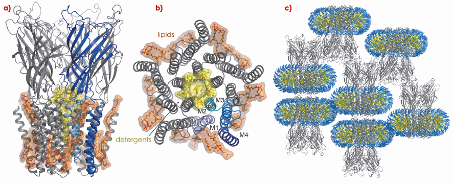
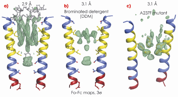
In the crystal structure of GLIC, some lipids were readily seen in the electron density in the vicinity of the TMD (Figure 113a,b). Modelling of the micelle “belt” around the TMD, compatible with the number of detergents bound in solution as determined from sedimentation experiments, resulted in a model entirely compatible with the crystal packing (Figure 113c). However, the big surprise in the crystal structure was that there was clear electron density in the pore itself. This could be unambiguously attributed to detergent molecules used to solubilise the protein (Figure 114a). N.B. The shape of the electron density does not change if one truncates the data to 3.1 Å.
 |
|
Fig. 113: General view of the crystal structure of the full-length ligand-gated bacterial ion channel GLIC: a) side view, b) top view c) packing interactions with modelled micelle in the crystal. |
To convince ourselves that the open pore conformation observed was not influenced by the binding of detergent inside it, we performed two other experiments: i) we collected diffraction data in the presence of brominated detergent, whose larger volume would suggest that all six detergent molecules seen in the original structure cannot bind simultaneously. We found an unaltered pore structure and a much weaker electron density for the detergents (Figure 114b); ii) we collected diffraction data for a mutant A237F where the side-chain phenylalanine was predicted (and observed) to protrude into the pore and exclude the detergent: again we found that the pore geometry is unaffected but that the density for detergent is now much weaker (Figure 114c).
 |
|
Fig. 114: Experimental electron density for the detergents in the pore: a) wild type protein with DDM (n-decyl-ß-D-maltopyranoside), b) wild type protein with brominated DDM, c) A237F mutant with DDM. |
Finally, we studied the stability of the open form in a fully hydrated and lipidic environment via a 20-ns long molecular dynamics simulation with about 200,000 atoms. The pore maintained an open form during that period of time.
There are many more questions to be answered for this family of proteins, which we are addressing through a combination of computational (molecular dynamics), structural (diffraction) and functional (electrophysiology) experiments on mutants and various different constructs.
References
[1] A. Taly et al., Nature Rev. Drug Discovery 8, 733-750 (2009).
[2] K. Brejc et al., Nature 411, 269-276 (2001).
[3] N. Unwin, J. Mol. Biol. 346, 967-989 (2005).
[4] A. Tasneem et al., Genome Biol 6, R4 (2005).
[5] R. Hilf and R. Dutzler, Nature 452, 375-379 (2008).
[6] N. Bocquet et al., Nature 445, 116-119 (2007).
Principal publication and authors
N. Bocquet (a), H. Nury (a), M. Baaden (b), C. Le Poupon (a), J-P. Changeux (a), M. Delarue (a) and P.-J. Corringer (a), Nature 457, 111 (2009).
(a) Pasteur Institute, Paris (France)
(b) Institut de Biologie Physico-Chimique, CNRS UPR 9080, Paris (France)


