- Home
- Users & Science
- Scientific Documentation
- ESRF Highlights
- ESRF Highlights 2010
- Electronic structure and magnetism
- Imaging multiferroic domains using X-ray polarisation-enhanced topography
Imaging multiferroic domains using X-ray polarisation-enhanced topography
Imaging ferroelectric and magnetic domains have largely remained two separate fields. Recent developments at beamline ID20 have led to a technique that provides topographic images of the spatial distribution of the magnetic domains in multiferroics, such as Ni3V2O8, while the ferroelectric domains are cycled through a hysteresis loop by an applied electric field. Ni3V2O8 is an example of a magnetoelectric multiferroic in which magnetism and ferroelectricity not only coexist, but are coupled together. These materials present an interesting opportunity for technological applications, but this is reliant on overcoming an outstanding challenge, namely understanding the evolution of the domain states in external applied magnetic fields. The work we present here is a first step in this direction.
In Ni3V2O8 there is a phase transition at T = 6.3 K to an incommensurate magnetic structure, that is accompanied by the onset of a finite spontaneous electric polarisation. Based on neutron diffraction, it was proposed that this phase corresponded to a cycloidal order of the spins on two different symmetry nickel sites – spine and cross-tie. Circularly polarised X-rays naturally couple to the handedness of spin cycloids, and through a complete Stokes polarimetry analysis of the scattered beam it is possible to identify information on magnetic structures that is typically inaccessible through neutron diffraction experiments. It was determined using just four reflections that, in fact, the moment on the cross-tie sites remains disordered, such that the onset of ferroelectricity is associated with the cycloidal ordering of the moments on the spine sites.
For our experimental setup, our sample of Ni3V2O8 was mounted such that the spin cycloid lay in the horizontal scattering plane, which resulted in the polarisation state of the scattered beam being strongly sensitive to the domain populations. The scattering process converts the incident circular polarisation state into a linear polarisation state inclined at 45 degrees to the scattering plane, described by the Stokes parameter P2. By determining the value of P2 at different points over the sample surface it is then possible to extract the domain populations.
 |
|
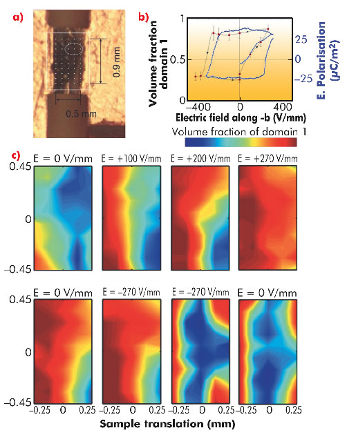
Fig. 82: a) Photograph of sample between the Cu electrodes forming a capacitor. b) Variation in the domain populations and the electric polarisation as a function of applied electric field. c) Topographic images of the domain population distributions as a function of the electric field. |
To establish the evolution of the domains as a function of field, the sample was initially cooled to 5 K with no voltage across the sample. The incident beam spot was then reduced in size and the sample rastered through the beam measuring the value of P2 at each spot and hence finding the percentage of left- and right-handed magnetic cycloidal domains. Then a voltage was applied whilst remaining at 5 K, before repeating the measurements of P2. Figure 82c reveals that this method allows us to resolve inhomogeneities in the domain populations with good resolution. The evolution of the domain populations is gradual, with the boundary between the two shifting as a function of applied electric field, as opposed to the nucleation of one domain within the other, which would lead to a more randomised distribution. It is also clear that the data reveals edge effects, where the domains become pinned close to the edges of the sample. By averaging the domain populations over the central unaffected area it is possible to extract a magnetic domain population hysteresis loop. On comparing this loop with electric polarisation measurements (Figure 82b), we find an excellent agreement, indicating that, by imaging the magnetic domains in multiferroic Ni3V2O8, we are in effect also imaging the ferroelectric domains.
The demonstration of the possibility to image the multiferroic domains in Ni3V2O8 opens up the prospect to use this technique to image domains in other related materials and even in operational devices. With the improvement in focussing offered by the ESRF Upgrade Programme, the spatial resolution of this technique will be reduced to 100 nm and below, at which point it will become possible to image not only the domains, but also the structure of the domain walls, the engineering of which ultimately determines the usefulness of any given material.
Principal publication and authors
F. Fabrizi (a,b), H.C. Walker (a,b), L. Paolasini (a), F. de Bergevin (a), T. Fennell (b,c), N. Rogado (d), R.J. Cava (d), Th. Wolf (e), M. Kenzelmann (f) and D.F. McMorrow (b), Phys. Rev. B 82, 024434 (2010).
(a) ESRF
(b) University College London (UK)
(c) ILL
(d) Princeton University (USA)
(e) Karlsruher Institut für Technologie (Germany)
(f) PSI (Switzerland)


