X-ray imaging
The set of highlights presented in this chapter shows, as usual, that the application of X-ray imaging techniques at the ESRF covers a very wide range of scientific domains: from biomedical applications and life sciences to materials, cultural heritage and environmental science. We are constantly striving to further improve the imaging capabilities, in particular lateral resolution, detection limits and speed of acquisition. Imaging is indeed one of the five flagships of Phase I of the ESRF Upgrade Programme. A set of upgrade beamlines (UPBL) are under construction, among which the NINA beamline composed of two independent branches: NI for nano-imaging, NA for nano-analysis. Its satellite building was inaugurated in October 2012 and hutches are currently being constructed. The ID19 beamline is also benefitting from a complete refurbishment partially carried out in conjunction with developments at ID17 and in the so-called paleontology facility. One of the numerous objectives of this refurbishment is to facilitate the transfer of samples between ID17 and ID19 to allow the combination of large field of view/medium resolution (ID17) with medium field of view/high resolution (ID19). This was achieved through the installation of four identical large sample stages (one at ID17, two at ID19, and also one at BM05) and the development of a standardised control system. Generally speaking, the combination of techniques, whether on a single beamline or on several, is an increasing trend. In the following examples, the elucidation of degradation processes affecting Van Gogh’s paintings was done by combining µXRF/µXRD at ID18F and µFTIR, µXRF/µXANES at ID21, and the study of geometrical carrier confinement within a single nanowire was carried out by collecting simultaneously optical luminescence and X-ray fluorescence.
Another general issue, illustrated by several of the following highlights, is the dose and the different approaches to limit possible radiation damage. At ID17, Zhao et al. implemented the so-called “EST based PCT” method, for equally sloped tomography based phase contrast X-ray tomography, which allows a simultaneous increase in resolution and decrease in the dose and acquisition time relative to conventional phase contrast tomography. Their work is illustrated by application to diagnosis of human breast cancers. Similar efforts to improve data acquisition and processing methods permitted further development of the X-ray grating interferometry technique at ID19, which offers a real trimodal (phase contrast, absorption contrast and scattering “dark-field” contrast) low-dose X-ray tomography.
Focusing the beam to micro and even nano probes makes dose and the consequent radiation damage even more critical. The use of a cryo-environment is a common solution, in particular for the study of biological samples. For the future NINA end-stations, the implementation of such environments is both mandatory and challenging. At ID21, different instrumental developments were recently implemented to improve the preparation and analysis of samples under cryoconditions. The study of TiO2 uptake in cucumber plants presented hereafter was carried out a few months ago at room temperature but would clearly have benefitted from these new cryo devices, judging from the success of similar recent experiments. The cryo-environment was also a necessity for the experiments of Deville et al. to follow the freezing of colloidal suspensions and subsequently observe the frozen microstructure, as done at ID19.
A room temperature alternative to cryo-microscopy was demonstrated in the article by Beerlink et al. where a native lipid bilayer membrane of less than 5 nm was imaged using an intense nanofocused beam in relatively low-dose conditions. This was made possible by positioning the sample a few millimetres downstream of the focal plane and by exploiting the contrast formed by the free space propagation of hard X-rays. Such an approach opens the way to many new investigations like membrane fusion, membrane phase transitions, and the transport of molecules through bilayers.
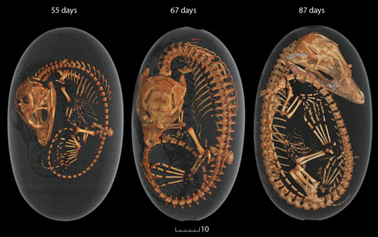
As in previous years, this chapter contains only a sub-set of many interesting results published during the year and obviously there are many omissions due to lack of space. In particular, two domains are not represented, which regularly use the X-ray imaging instruments: palaeontology (e.g. fossil bone 3-D virtual histology [Sanchez et al., Microscopy and Microanalysis 18, 1095-1105 (2012)]) and evolutionary biology (e.g. in ovo 3-D embryology on crocodiles, Figure 55); and Earth sciences, where the micrometric resolution and imaging capabilities are regularly associated to X-ray absorption spectroscopy in order to identify and localise different speciations of key elements such as iron, and consequently monitor fundamental redox processes [Borfecchia, J. Anal. At. Spectrom. 27, 1725 (2012); Beck, Geochimica et Cosmochimica Acta 99, 305 (2012)].
2013 will be a fundamental year for X-ray imaging at the ESRF. Work will continue for the phase I projects of UPBL4-NINA and the ID19/ID17/paleontology refurbishment with various steps of implementation and commissioning being scheduled. Discussions will also start regarding phase II. While phase I was oriented towards improved resolution (in particular nanoscopy), phase II could be the opportunity to explore temporal resolution. Hence the name of the workshop, satellite to the 2013 Users meeting: “X-ray cinematography with the new coherent source”.
M. Cotte


