- Home
- Users & Science
- Scientific Documentation
- ESRF Highlights
- ESRF Highlights 2015
- Structure of materials
- Brilliant Synchrotron radiation and electron microscopy – a powerful combination for challenging structure analyses
Brilliant Synchrotron radiation and electron microscopy – a powerful combination for challenging structure analyses
Extremely brilliant microfocused synchrotron radiation enables the extension of “standard” single-crystal structure determination to samples smaller than one micrometre. In combination with transmission electron microscopy, novel compounds in inhomogeneous reaction products can be discovered and accurately characterised. Taking this information into account allows one to develop efficient syntheses.
Structure analyses of compounds present in heterogeneous samples can be demanding, especially if metastable phases or the products of precursor reactions do not exhibit crystals with sizes required for conventional structure analysis. Powder X-ray diffraction is, in many cases, insufficient for the precise and accurate characterisation of such materials with unknown structure types. Despite recent advances to overcome the problem of dynamical diffraction, electron crystallography still cannot compete with the accuracy of single-crystal structure analyses.
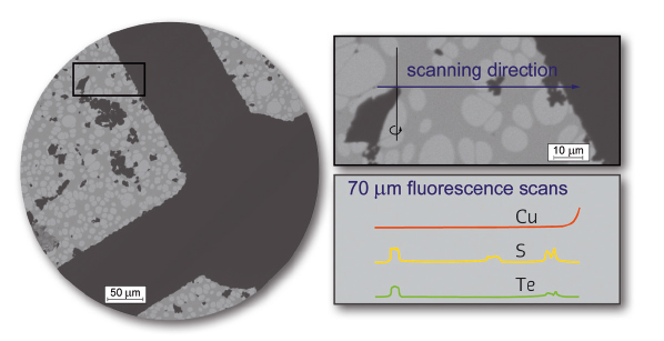
The very stable beam at beamline ID11 can be focused to diameters of less than one micrometre by refractive lenses. This enables precise X-ray structure analyses of microcrystals which can be centred by detecting their characteristic X-ray emission while moving the sample in the beam (fluorescence scans, Figure 28). However, it would be extremely tedious to identify a good crystallite of a new compound that occurs as a side phase in heterogeneous materials. Transmission electron microscopy (TEM), on the other hand, is a reliable tool for the discovery of such new phases and the determination of their unit-cell metrics by electron diffraction and their chemical composition by energy-dispersive X-ray spectroscopy. TEM and microdiffraction were efficiently combined by using microcrystalline samples on carbon-coated copper grids. Crystallites were pre-characterised by TEM and re-located at the beamline by fluorescence scans so that diffraction data could be acquired.
 |
|
Fig. 28: TEM image of the selected crystallite on a copper grid (left, scanned region marked by black box) and an enlarged image of the measured tip of the crystallite (0.3 × 0.5 × 1 μm3) with centre of rotation (top right), and a schematic representation of fluorescence line scans of Cu, Te and S (bottom right). The copper crossbar functions as a “landmark”; a side-phase visible in the centre of the scan contains no Te, whereas the crystals on the right side of the scans are located too close to the copper bar to be measured independently. |
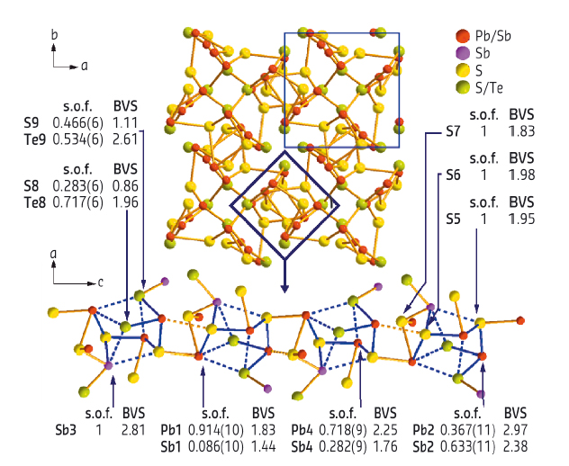
As an example, the very unusual compound Pb8Sb8S15Te5 was found in quenched samples with the nominal composition “Pb5Sb4S6Te5” that contained a mixture of PbTe, Sb2Te, and an unknown quaternary phase. The latter’s metrics and approximate composition as determined by TEM methods revealed the presence of a new sulfide telluride. The lack of single crystals and strongly overlapping broadened reflections in powder diffraction patterns impeded further characterisation. The tip of a pre-characterised quaternary crystallite (ca. 0.3 x 0.5 x 1 mm3) in a suitable and accessible position on the grid was analysed at ID11. The resulting dataset turned out to be precise enough for a detailed description of the structure, which was refined including twinning around [110] and additional inversion twinning. Mixed site occupancies as well as anisotropic displacement parameters could be elucidated as in any high-quality X-ray analysis of macroscopic single crystals. Pb8Sb8S15Te5 crystallises in space group P41 (a = 8.0034(4) Å, c = 15.0216(5) Å) and exhibits an intriguing structure with chain-like fragments in combination with heterocubane-like entities (Figure 29). The structure model was further confirmed by the excellent match of simulated and experimental high-resolution TEM images of the very crystallite. Furthermore, the detailed knowledge of the structure and exact composition led to the subsequent synthesis, optimisation and thermoelectric characterisation of phase-pure material.
 |
|
Fig. 29: Crystal structure of Pb8Sb8S15Te5 as obtained by microfocus diffraction with bond valence sums and site occupancies; characteristic chain-like unit with heterocubane-like building blocks (bottom). |
This combined TEM and microdiffraction approach avoids the once tedious and time-consuming “trial and error” structure analysis of microcrystalline heterogeneous samples and opens new perspectives in solid-state synthesis, which can now follow the full structural characterisation instead of being a requirement for it. Selecting specimens for X-ray diffraction by TEM can also be applied to intergrown domains in composite materials and other fields of materials science (for further examples, see refs. [1,2]). In addition, this strategy might also be applied to the study of reaction pathways and intermediates.
Principal publication and authors
Discovery and structure determination of an unusual sulfide telluride through an effective combination of TEM and synchrotron microdiffraction, F. Fahrnbauer (a), T. Rosenthal (b), T. Schmutzler (a), G. Wagner (a), G.B.M. Vaughan (c), J.P. Wright (c) and O. Oeckler (a), Angew. Chem. Int. Ed. 54, 10020-10023 (2015); doi: 10.1002/anie.201503657.
(a) Faculty of Chemistry and Mineralogy, Leipzig University (Germany)
(b) Department of Chemistry, LMU Munich (Germany)
(c) ESRF
References
[1] F. Fahrnbauer et al., J. Mater. Chem. C 3, 10525 (2015).
[2] D. Durach et al., Inorg. Chem. 54, 8727 (2015).


