- Home
- Users & Science
- Scientific Documentation
- ESRF Highlights
- ESRF Highlights 2017
- X-ray nanoprobe
- A biological strategy for pre-stressing crystalline calcite lenses
A biological strategy for pre-stressing crystalline calcite lenses
A biostrategy has been presented for strengthening and toughening the otherwise brittle calcite optical lenses found in the brittlestar Ophiocoma wendtii. This intriguing process employs coherent nanoprecipitates to induce compressive stresses on the host matrix, functionally resembling the Guinier–Preston zones known in classical metallurgy, though produced at ambient conditions without heating and quenching.
Various functional materials are formed via the process of biomineralisation [1] and serve specific functions [2]. Fascinatingly, these materials are formed from limited bioavailable elements, without the possibility of heating and quenching, as is common in man-made counterparts. Instead, organisms utilise various alternative biostrategies.
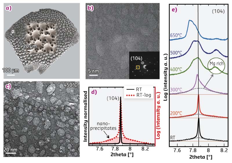
A new biostrategy has been revealed in the Ophiocoma wendtii brittlestar, which has a unique micro-lens system made of calcite spread over its dorsal armplates (Figure 82a). Coherent nanoprecipitates revealed in the matrix were found to induce comprehensive compressive stresses on the host single crystal, akin to the Guinier–Preston zones known in classical metallurgy. These nanoprecipitates are rich in magnesium, which substitutes calcium in the lattice and leads to the shrinkage of lattice parameters, and this is the source of compressive stresses on the host matrix. This engineering technique strengthens and toughens ceramic materials, similar to that employed in tempered glass and prestressed concrete.
Utilising high-resolution powder diffraction on ID22, it was discovered that magnesium-rich nanodomains exist within the single crystal and are coherent within the matrix. This is concluded from the hump that appears at the base of each of the diffraction peaks prior to annealing (Figure 82d). The nanoprecipitates were also observed by aberration-corrected transmission electron microscopy (TEM), which also reveals their relatively low electron density and coherent interfaces with the matrix (Figure 82b). Once the sample is heated, these domains grow in size unless coherency is lost at the interface and the strain is released. This behaviour was observed both by in situ heating in the TEM as well as in situ heating on ID22 (Figure 82c, e). In the latter, it was found that the diffraction peaks from the nanophase emerge only after 400°C and are shifted to higher 2-theta positions as compared to those of the matrix diffraction peaks, due to a higher magnesium concentration and hence smaller lattice parameters (Figure 82e).
 |
|
Fig. 82: a) SEM image showing an entire dorsal armplate and a higher magnification inset. b) TEM image of a thin section from a lens, revealing brighter nanodomains and their coherence with the matrix, although the FFT pattern is that of a single crystal (inset). c) Bright-field TEM image acquired during in situ heating at 450°C, revealing the temperature-induced growth of the nanodomains. d) The (104) diffraction peak comparing linear (black) and logarithmic (red) intensity scales, and revealing the presence of nanodomains at the base of the diffraction peak. e) Evolution of the (104) diffraction peak with temperature. At 400°C, a distinct broad diffraction peak appears owing to the loss of nanoprecipitate coherence. |
Sub-micron diffractometry on ID13 was used to map the variation in d-spacings and SAXS signal of a single lens. Both maps revealed structured regions slightly beneath the surface appearing in curved lines parallel to the surface (Figure 83a, b). These layers were also visualised via nano-computed tomography (nanoCT) measurements obtained on ID16B, revealing alternating layers of density, indicated by the brightness variation in phase contrast (Figure 83c). The alternating densities are likely due to the different concentrations of magnesium-rich nanoprecipitates, which coincides with the diffraction-mapping features.
 |
|
Fig. 83: a) D-spacings map of a single lens area of the (214) reflection. b) SAXS intensity map of the same lens (~2.5−16.5 nm). c) A virtual slice within a single lens produced by nanoCT, revealing alternating density layers probably owing to varying nanoprecipitate content. |
Brittlestar lenses are formed via an amorphous precursor phase that crystallises at room temperature. This amorphous phase is supersaturated with magnesium as compared to the crystalline calcite, which drives the formation of segregated nanoprecipitates rich in magnesium after or during crystallisation. The nanometric size of the precipitates enables them to bear high tensile stresses while exerting compressive stress on the host matrix via the coherent interfaces. This is very similar to what is known in conventional metallurgy as Guinier-Preston zones, however, in contrast, biology forms this microstructure at ambient conditions without the need to heat and quench.
Principal publication and authors
Coherently Aligned Nanoparticles Within a Biogenic Single Crystal: Biological Prestressing Strategy, I. Polishchuk (a), A.A. Bracha (a), L. Bloch (a) , D. Levy (a), S. Kozachkevich (a), Y. Etinger-Geller (a), Y. Kauffmann (a), M. Burghammer (b), C. Giacobbe (b), J. Villanova (b), G. Hendler (c), S. Chang-Yu (d), A.J. Giuffre (d), M.A. Marcus (e), L. Kundanati (f), P. Zaslansky (g), N.M. Pugno (f), P.U.P.A. Gilbert (d), A. Katsman (a) and B. Pokroy (a), Science 358, 1294-1298 (2017); doi: 10.1126/science.aaj2156.
(a) Department of Materials Science and Engineering and the Russel Berrie Nanotechnology Institute, Technion-Israel Institute of Technology (Israel)
(b) ESRF
(c) Natural History Museum of Los Angeles County (USA)
(d) Departments of Physics, Chemistry, Geoscience, University of Wisconsin-Madison, (USA)
(e) Advanced Light Source, Lawrence Berkeley National Laboratory, California (USA)
(f) Laboratory of Bio-Inspired & Graphene Nanomechanics, University of Trento, (Italy)
(g) Department for Restorative and Preventive Dentistry, Centrum für Zahn-, Mund- und Kieferheilkunde, Charité-Universitätsmedizin Berlin, (Germany)
References
[1] H. A. Lowenstam and S. Weiner, On Biomineralization, Oxford University Press (1989).
[2] J.W.C. Dunlop and P. Fratzl, Annu. Rev. Mater. Res. 40, 1-24 (2010).


