- Home
- News
- General News
- A unique atlas of...
A unique atlas of the human heart: from cells to the full organ
17-07-2024
Scientists led by the University College London (UCL) and the European Synchrotron (ESRF), have, for the first time, imaged two whole human adult hearts, one healthy and one diseased, down to the cellular level in 3D, using an innovative X-ray technique called Hierarchical Phase-Contrast Tomography (HiP-CT). This new atlas of the heart can potentially lead to medical applications. The results are published in Radiology.
Share
To study the human heart, researchers typically use clinical imaging techniques such as Ultrasound, Computed Tomography (CT) and Magnetic Resonance Imaging (MRI). While these methods are effective for diagnosing cardiovascular disease, they do not provide detailed structural changes across the different scales within the heart. For higher resolution, histology is required, which involves slicing donor organs into sections. Although this method offers more detailed information, it significantly limits the field of view.
Now a new synchrotron X-ray imaging technique, called HiP-CT, overcomes these limitations by providing a comprehensive and detailed 3D view of the entire adult human heart. “HiP-CT provides a global view of whole donor organs at unprecedented resolution, bridging the gap between traditional imaging and histology,” says Professor Peter Lee of UCL, HiP-CT project lead.
 |
|
Click on the image to view the video: Multidimensional Analysis of the Human Heart in Health and Disease using Hierarchical Phase-Contrast Tomography (HiP-CT). Credit Brunet et al. Radiology. |
A team led by UCL and the ESRF, in collaboration with the Wellcome Sanger Institute, Siemens Healthineers, Great Ormond Street Hospital, Hannover Medical School, the Aachen Medical University, the Helios University Clinic Wuppertal, the University Medical Center of the Johannes Gutenberg-University Mainz, and the Laboratoire d’Anatomie des Alpes Francaises (LADAF) has imaged two entire adult hearts, one healthy and one diseased, using the HiP-CT imaging technique on beamline BM18 at the ESRF. “BM18 is currently the only place in the world where complete human organs can be imaged with such a high level of contrast, and we are still quite far from the limits of the beamline capabilities. The main limiting factor is the processing of the very large data produced by HiP-CT”, explains Paul Tafforeau, ESRF scientist.
 |
|
Joseph Brunet, researcher at UCL and ESRF visiting scientist and Hector Dejea I Velardo, junior scientist member of the Humain Organ Atlas Porject, during the experiments at the ESRF, the European Synchrotron, at BM18 beamline. Using an advanced synchrotron technique called HiP-CT, scientists have imaged, for the first time, two whole human hearts at 20-micron resolution, zooming in to 2-micron details in 3D. Credit: ESRF/Stef Cande. |
One of the major advantages of this technique is that it achieves a full 3D view of the organ at a resolution of 20 microns, which is around 20 times better resolution than a clinical CT scanner. In addition, it can then zoom in to cellular level at 2-microns (200 times better), achieving histological resolution without cutting the sample. This technique images whole organs hierarchically, revealing details and connections that were previously unknown.
 |
|
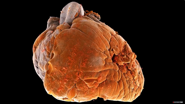
Using an advanced synchrotron technique called HiP-CT, scientists have imaged, for the first time, two whole human hearts at 20-micron resolution, zooming in to 2-micron details in 3D. Credit: Brunet et al., Radiology. |
Whilst there have been synchrotron studies on whole fetal and small animal hearts before, these have always been at a tiny scale, 1 or 2 cm in diameter. In this new research, scientists have successfully imaged adult hearts of 14 cm in diameter.
Phase contrast imaging differs from conventional X-ray imaging, which relies on X-ray absorption, by exploiting the refraction of specific types of X-rays passing through tissues, rather than relying solely on X-ray absorption. It results in images with dramatically higher contrast and resolution, particularly beneficial for visualising soft tissues and fine structures, as found in the heart, without the need for staining with a contrast agent.
HiP-CT showed its capacity for high spatial resolution, multi-scale, cardiac imaging ex-vivo, revealing histologic-level detail of the myocardium (muscle cells), valves, coronary arteries and cardiac conduction system (the electrical wiring triggering heart contraction) across length-scales. “The first time you see the heart with HiP-CT, it is quite surprising, as it clearly shows soft tissue not typically visible with conventional X-ray imaging”, says Joseph Brunet, researcher at UCL and ESRF visiting scientist, corresponding author of the publication.
 |
|
3D cinematic renderings of the control and diseased heart in anatomic orientation. Epicardial fat has been removed digitally to show course of the major coronary arteries plus detail of smaller arteries penetrating into the myocardium which are not typically seen on clinical CT. In the control (A), the coronary arteries remain close to the epicardial surface while in the diseased heart (B) they are lifted away by epicardial fat increasing the perfusion distance between the major coronaries and the myocardium. Segmentations and high resolution detail of coronaries in the diseased heart are also shown in Figure 5. Credit: Brunet et al., Radiology. |
Towards new arrhythmia treatments
A significant feat of the study is the detailed imaging of the cardiac conduction system, which generates and transmits the electrical signals that drive the heart muscle’s pumping action.
The virtual sectioning of the conduction system provided scientists with information on fatty infiltration, vascular supply, and pathways between the cardiac nodes and adjacent structures. This level of detail on the entire adult heart conduction system was previously unattainable with existing imaging techniques.
“There is an enormous potential to inspire new treatments using this technique”, explains Professor Perry Elliot, Director of the Institute of Cardiovascular Science, UCL. An example of this is arrhythmia: “With today’s technology, having an accurate interpretation of the anatomy underlying arrhythmia is very difficult”, adds Professor Andrew Cook, heart anatomist at UCL and second author on the paper.
“We believe that our findings will help researchers understand the onset of cardiac rhythm abnormalities and also the efficacy of ablation strategies to cure them”, he says. In particular, the results have shown that scientists can determine differences in the thickness tissue and fat layers located between the outer surface of the heart and the protective sac surrounding the heart. This information could be relevant when treating arrhythmia (irregular rhythm).
In addition to arrhythmia, HiP-CT can also shed light on other cardiovascular diseases. Anatomical studies using HiP-CT are currently underway to investigate congenital heart defects such as single ventricle disease. “The practical flexibility and very detailed data generated, makes HiP-CT ideal for understanding complex alterations to organ anatomy in the course of disease” adds Prof. Danny Jonigk, Chair of the Institute of Pathology at RWTH Aachen.
For the team, the next step is to increase the number of samples and to “continue exploring the structural architecture of the heart in health as well as in disease, with the aim of developing innovative diagnostic and treatment strategies”, concludes Cook.
This work contributes to the Human Organ Atlas project, which aims to establish an open science image database of all human organs in health and disease. To explore this data yourself visit human-organ-atlas.esrf.eu, or for more information on the Human Organ Atlas project see mecheng.ucl.ac.uk/HOAHub/ and videos at bit.ly/HiP-CT-Heart.
The project is co-funded by the Chan Zuckerberg Initiative (CZI).
Reference:
Brunet, J. et al, Radiology. https://doi.org/10.1148/radiol.232731
Top image: 3D cinematic renderings of the control and diseased heart in anatomic orientation. Credi: Brunet et al., Radiology.
partners


European Synchrotron Radiation Facility - 71, avenue des Martyrs, CS 40220, 38043 Grenoble Cedex 9, France.