- Home
- News
- Spotlight on Science
- Three-dimensional...
Three-dimensional nano-imaging of the brain
20-08-2018
X-ray nanoholotomography allows large-scale, label-free, direct imaging with isotropic voxel sizes well below 100 nm. It is especially attractive for the non-destructive hierarchical imaging of human tissues as it avoids physical slicing and therefore complements well-established histology and electron microscopy.
Share
Pathologists generally examine micrometre-thin tissue slices by means of optical microscopy in order to diagnose potential diseases. More recently, high-resolution hard X-ray microtomography (µCT) has been applied to complement histology by adding the third dimension [1,2]. The lateral spatial resolution of the optical micrographs of the histological slices, however, has been an order of magnitude better than for µCT. Through the combination of X-ray optics and the phase contrast mode at the nano-imaging beamline ID16A, the spatial resolution has been greatly improved, reaching deep into the nanometre range, i.e. well beyond the optical limit. The embedding of the human brain tissue into paraffin, as is common practice in clinics, results in an amazing contrast, allowing for the identification of numerous anatomical features of nanometre size, see below. Consequently, the imaging technique, which has been developed and implemented by the team at ID16A, can be regarded as a milestone in the establishment of nanoanatomy as a fundamental chapter of nanomedicine.
The term holotomography coined in 1999 [3] relates to the in-line Gabor holography introduced in 1948 [4] and stems from the combination of holographic and tomographic reconstructions. The term holography was introduced by D. Gabor to indicate that the method records the complete wave front, i.e. amplitude and phase. Therefore, the holographic reconstruction, which is done numerically, recovers both the real and imaginary parts of the refractive index. For in-line holography, a partially coherent beam interferes with itself so that no separate reference beam is required. Thus, in-line holography is considered the poor man's holography, but in fact it is the original and the oldest one.
Hard X-ray nanoholotomography (XNH) is regarded as non-destructive because it does not require physically slicing the specimen and multiple measurements exhibit the same result, even after long X-ray exposure. The 180° acquisition and tomographic reconstruction creates a virtual three-dimensional model without destroying the original object, here the selected human brain tissues were treated according to the standard protocols in pathology for a wide variety of daily diagnoses.
XNH has numerous advantages with respect to the established techniques. First, the penetration depth of the hard X rays is much larger than for visible light and electrons, which makes mechanical slicing obsolete. Second, the employment of local phase tomography allows for hierarchical imaging (see Figure 1). This multiscale approach includes a fast overview scan of a volume close to one cubic millimetre in order to select a region of interest. This region can be subsequently investigated with voxel volumes three orders of magnitude smaller. Third, after XNH, the same tissue can be used for the well-established histological analysis.
 |
|
Figure 1. Hierarchical brain imaging down to the true nanometre scale. XNH bridges the resolution gap in three-dimensional imaging between µCT and electron microscopy with tissue ablation. |
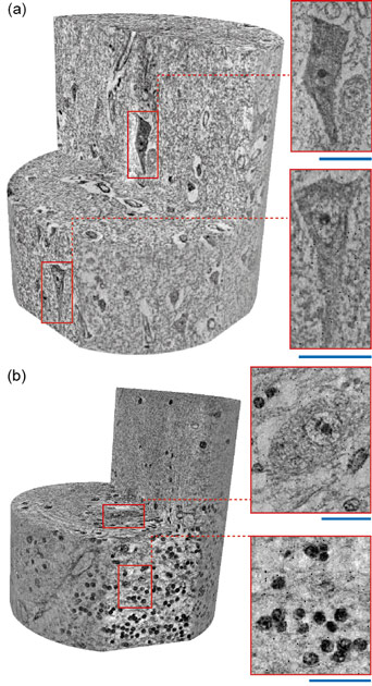
Figure 2 represents two local tomograms of human brain tissues, namely neocortex and cerebellum. The volume rendering of the brain tissue after clinically-relevant tissue preparation but without cutting highlights the electron microscopy-like data quality at 100 nm voxel length. The virtual cutting planes illustrate the presence of cellular and even subcellular features, which include cell somata, dendrites, nuclear envelopes, nucleoli, and structures within the nucleus. The scale bars correspond to a length of 20 μm. It is known that the desired spatial resolution restricts the volume accessible. Local tomography approaches allow for the combination of volumes measured separately within the same object, so that a significant portion of the human brain can be imaged. As the handling of big data advances, visualisation of the entire brain with nanometre resolution could become available during the next decade.
Principal publication and authors
Hard X-ray nanoholotomography: large-scale, label-free, 3D neuroimaging beyond optical limit, A. Khimchenko (a), C. Bikis (a), A. Pacureanu (b), S.E. Hieber (a), P. Thalmann (a), H. Deyhle (a), G. Schweighauser (c), J. Hench (c), S. Frank (c), M. Müller-Gerbl (d), G. Schulz (a), P. Cloetens (b), and B. Müller (a), Advanced Science 5, 1700694 (2018); doi: 10.1002/advs.201700694.
(a) Biomaterials Science Center (BMC), Basel (Switzerland)
(b) ESRF
(c) Institute of Pathology, Basel (Switzerland)
(d) Musculoskeletal Research Group, Basel (Switzerland)
References
[1] A. Khimchenko et al., NeuroImage 139, 26 (2016).
[2] S.E. Hieber et al., Scientific Reports 6, 32156 (2016).
[3] P. Cloetens et al., Applied Physics Letters 75, 2912 (1999).
[4] D. Gabor, Nature 161, 777 (1948).


