- Home
- Users & Science
- Scientific Documentation
- ESRF Highlights
- ESRF Highlights 2002
- X-ray Imaging
- Synchrotron-radiation Microtomography
Synchrotron-radiation Microtomography
Review by P. Cloetens and J. Baruchel (ESRF)
Microtomography is the three-dimensional (3D) reconstruction, via a numerical process, of the structure of a sample from a set of two-dimensional images, with a resolution of a few micrometres or less. Synchrotron radiation microtomography is now routinely used on several beamlines to perform investigations on a wide range of topics. Like other imaging techniques, it can provide information on samples from physical, medical, materials science and engineering subjects. Furthermore, the technique is also able to innovate in new areas like geophysical, environmental, paleontological and biological studies. This technique occupies now most of the beam time on ID19, and a substantial fraction on ID15, ID17 and ID22 and demand is clearly increasing. The three-dimensional data provided by microtomography is essential for the understanding of a wide range of phenomena [1,2].
When considering the present situation, we could be surprised that this use was not foreseen at the early stages of the ESRF, only 15 years ago (the "Red book"). The reason resides in the fact that microtomography not only requires the very brilliant sources provided by the ESRF, but also adapted detectors and very high computer power both in terms of calculation speed and memory. Indeed one 3D image represents about 2 Gigabytes of data, which is presently recorded in, typically, 10 minutes. This has to be compared with the total central memory foreseen for the ESRF at the time of the Red book, i.e. 3 Gigabytes. The effort devoted at the ESRF to implement high-resolution X-ray detectors based on a CCD camera exhibiting simultaneously a high dynamic range and a fast readout (the FReLoN camera), together with the general progress of the computers, when coupled with the ESRF beams, resulted in the microtomographic possibilities we have today.
Intensity is the first quantitative feature that has made progress in microtomography possible. It allows either the use of a monochromatic beam, which makes the quantitative evaluation of the results possible, and eliminates artefacts from the reconstructed image, or the implementation of fast tomography, which has been for instance used to follow the sintering of copper spheres.
In addition, when working at the ESRF, the physical effect that leads to image formation is not only the attenuation of the beam, essentially through photo-electric absorption, but also its phase shift suffers on going through a specimen. Contrast from phase features can be obtained with high instrumental simplicity through mere propagation over finite distances. A quantitative 3D reconstruction of the phase shift (corresponding to the local electron density) is now routinely achieved, based on images recorded at several distances ("holotomography") [3].
The ESRF is the place where the most significant progress of the relatively young synchrotron radiation microtomography technique has been achieved. This is why representative results indicating the developments that have emerged can be found in previous issues of the ESRF Highlights. Among them let us mention:
- the quantitative analysis of bones, which showed the actual effect of drugs used to treat osteoporosis,
- the observation of Uranium particles from Chernobyl soils, where cavities and channels reflect the formation of fission products during reactor operation,
- the holotomographic investigations of semisolid alloys, which allows one to visualise phases which display less than 2% difference in density, and of the seed of Arabidopsis thaliana, a favourite among plant geneticists, which is imaged in the damp state, without any specimen preparation.
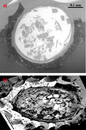
A recent result showing the state of the art is the one on charophytes, which are members of the green algae group. They are supposed to play an important role in the evolution towards vascular plants. Their fertile elements are well conserved in a calcified state (typically calcite) and fossils are often found in limestones and marls, deposited in brackish or freshwater environments. The analysed fossil consists of an inner structure and an outer protective sheath, formed by the growth of elongated spiral cells around the core. The observation of contrast between the sample that is mineralised in calcite and the limestone bulk necessitates a complete holotomographic reconstruction (Figure 89).
 |
|
|
The future progress in microtomography is foreseen along various ways, which include the combined technique approaches, fast tomography and magnified tomography.
The possibilities of combined approaches, in which tomography cooperates e.g. with topography ("topo-tomography") or fluorescence have already been implemented and demonstrated in a few cases. In this last case, the local character observed by fluorescence implies the use of a microbeam, and tomography is obtained by recording a scanned image for each orientation, and recording the fluorescence associated with each point in the map. The chemical state of an element can, alternatively, be visualised through the combination with X-ray absorption near-edge structure (XANES).
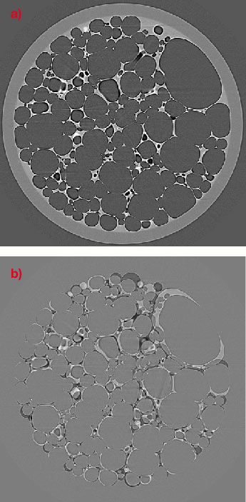
Fast tomography is very important to be able to follow evolving phenomena. One example application is given in Figure 90, which shows a liquid foam (a) and its evolution over 35 minutes through the subtraction of two successive images (b): this last image indicates that coarsening tends to increase the volume of the large bubbles at the expense of the small ones.
 |
|
|
|
|
|
The emergence of magnifying devices is another exciting area, which allows us to perform 3D images with sub-micrometre spatial resolution. Presently there is no X-ray magnification of the image so the resolution limit of microtomography is set by the detectors used. A very promising approach allowing this magnification is based on the use of curved mirrors and multilayers in the Kirkpatrick-Baez (KB) geometry. X-ray focusing to about 102 µm2 spot size with 5.1011 ph/s at a photon energy of 20 keV has been very recently demonstrated using a KB device. A magnified image is obtained when the detector is placed further from the specimen than the focus to sample distance (projection microscopy). The extension to microtomography is not straightforward in a routine mode, because it implies mechanical precision and stability, which are at the very limit of what is presently achievable, and therefore requests substantial technical developments.
Let us note, to conclude, that the use of microtomographic data is evolving. In addition to its industrial interest, the major scientific point to note is the emergence of imaging from taking 'snapshots' or 'videos' to extracting quantitative data from images suitable for model building or evaluation.
References
[1] Developments in X-Ray Tomography III, U. Bonse, editor, Proceedings of SPIE 4503 (2002).
[2] P. Cloetens, W. Ludwig, E. Boller, F. Peyrin, M. Schlenker, and J. Baruchel. Image Anal. Stereol. 21(Suppl 1), S75-S85 (2002).
[3] P. Cloetens, W. Ludwig, J. Baruchel, D. Van Dyck, J. Van Landuyt, J. P. Guigay, and M. Schlenker, Appl. Phys. Lett., 75:2912-2914 (1999).


