- Home
- News
- Spotlight on Science
- Ultra-fast processes...
Ultra-fast processes imaged with picosecond hard X-ray flashes
10-10-2014
Ultra-fast processes such as shock waves or crack propagation in solid matter can now be studied by direct space imaging thanks to single-bunch imaging techniques. The flash of X-ray light from a single bunch of electrons in the storage ring lasts only a few hundred picoseconds but it can be exploited to gain access to a new time domain. This technique is demonstrated here by the examples of a car fuse melting and a pane of glass shattering. The outlook for ultra-fast imaging of this kind is promising as Phase II of the ESRF Upgrade Programme will foster single-bunch imaging techniques even further with its increase of coherent photon flux at higher energies.
The potential of hard X-ray imaging to tackle scientific challenges in fields such as materials science can be substantially increased when time is accessible as a new dimension. Until now, the temporal resolving power of X-ray imaging at synchrotron light sources was generally given by the shortest exposure time accessible via the detector, i.e. down to a few microseconds [1]. With the possibility of using only the light from a single-bunch of electrons in the storage ring, this value drops by four orders of magnitude down to a few hundred picoseconds [2].
 |
|
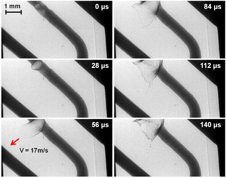
Figure 1. Time series showing pore formation in an electric wire leading to the rupture of a fuse. The pictures were acquired via single-bunch imaging at beamline ID15A. A movie includes this series: breaking of the fuse. |
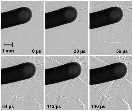
For the first proof-of-concept experiments, the ESRF was operated in a dedicated single-bunch beam mode. With 10 mA in the single bunch, the measured duration of the corresponding X-ray flash was 140 ps (FWHM). The revolution frequency of the single bunch is determined by the storage-ring circumference (844 m): 355.042 kHz, corresponding to 2.81 µs which is “long” enough to allow separation of the light flashes by the commercial CMOS-based cameras installed at the ESRF. In this special mode of operation, we were able to capture a car fuse breaking under high current using beamline ID15A. With such unprecedented time resolution, we observed an expanding pore inside the molten metal wire while it ruptured the connection across the fuse (Figure 1). The images are of remarkably high quality in terms of sharpness and contrast. A metal pore lamella, which is expanding at 17 m/s, is visible, while the images show no blurring (there is no ‘double image’ visible). In a parallel experiment, ID19’s superb coherence properties were used to track crack propagation in a piece of glass that was being shattered though the impact of an accelerated bolt (Figure 2). Considering that cracks can propagate in solid matter with ultrasonic speed, the acquired images are of excellent quality in terms of sharpness and contrast.
 |
|
Figure 2. Time series recording crack propagation in a glass plate initiated by an accelerated bolt. The pictures were acquired via single-bunch imaging at beamline ID19. A movie includes this series: crack propagation in glass. |
Today’s X-ray imaging experiments with ultimate time resolution on storage rings are mainly limited by the available source brilliance and detector performance both in terms of spatial and time resolution. With the ESRF Upgrade Programme entering Phase II, an increase of about two orders of magnitude in source brilliance, which translates directly into a corresponding increase of the coherent fraction of X-ray photons, can be expected from the new machine (see: ESRF Upgrade Programme Phase II (2015‐2019) White Paper). This will trigger the evolution of ultrafast imaging from a niche application towards a routine tool in materials science and also leads the way to other exciting opportunities for future materials science applications such as the realistic prospect of single-bunch diffraction and single-bunch spectroscopy (see: ESRF sends shockwaves (and an X-ray pulse) through iron to prove a new technique). The expected performance of new near-diffraction limited light sources thus constitutes a big step towards new applications of single-bunch diffraction, spectroscopy and imaging for materials science.
Principal publication and authors
Exploiting coherence for realtime studies by single-bunch imaging, A. Rack (a), M. Scheel (b), L. Hardy (a), C. Curfs (a), A. Bonnin (c), H. Reichert (a), Journal of Synchrotron Radiation 21, 815-818 (2014).
(a) ESRF
(b) Synchrotron Soleil, Gif sur Yvette (France)
(c) Paul-Scherrer Institute, Villigen (Suisse)
References
[1] A. Rack et al., Applied Optics 52, 82122 (2013).
[2] S.N. Luo et al., Rev. Sci. Instrum. 83, 073903 (2012).
Top image: 140 ps snapshot of a fuse rupturing.


