- Home
- News
- Spotlight on Science
- Coarsening and break-up...
Coarsening and break-up revealed by X-ray microtomography of phase-separated glasses
23-12-2014
A team from ESPCI, CNRS and Saint-Gobain studied the microstructure formation in a molten silicate glass during high-temperature heat treatment using X-ray microtomography at beamline ID19. They observed the glass phase separate into two phases. Initially the phases had an interpenetrating geometry, but later on one phase began to fragment and progressively became isolated droplets.
In their molten state, some silicate glasses spontaneously phase-separate: they form domains of different compositions [1, 2]. These domains can take different shapes, such as droplets or interconnected channels. The physics is then very similar to that of an emulsion of oil and water, with the microstructure gradually coarsening to reduce the interfacial energy. In the case of a silicate glass, the liquid state is obtained at temperatures generally higher than 700°C, and by cooling it below this temperature, one can “freeze” the system. The domains formed in the liquid state then become a microstructure in the solid, and can be used to tune some physical properties. Using X-ray tomography, we were able to visualise the formation of this microstructure in a barium borosilicate glass in three dimensions (3D), and observe the effect of the time spent in the liquid state.
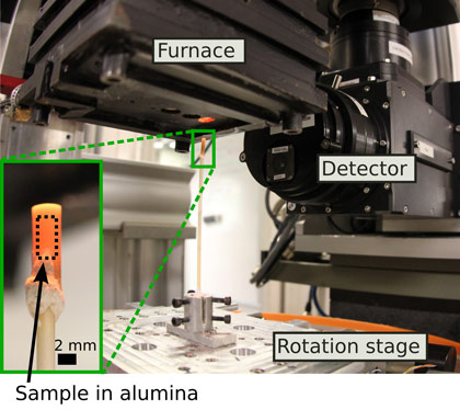
X-ray tomography comprises the reconstruction of 3D images of the absorption of a sample from several hundreds of projections. Fast scans are possible at beamline ID19 by the high intensity flux together with a setup suited to ultrafast data acquisition. Moreover, a dedicated furnace was used such that the same sample can be heated for a certain time at a given temperature then quenched to be scanned, then heated again, and so on (Figure 1).
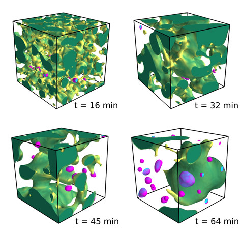
After image processing, one can observe the intricate geometry of one of the two phases (Figure 2). At the beginning of the heat treatment, the system is bicontinuous, i.e., there is only one domain for each phase, and these domains are interpenetrating. The geometry is similar to a sponge, where there is only one “solid” part, that is contiguous (otherwise it would collapse), and only one “void” part (otherwise the water could not fill it all). One can in fact produce a “glass sponge” by leaching one of the two separated phases with acid, to produce porous glass membranes for instance.
During the heat treatment, we were able to measure the increase in typical size of the domains that occurs in order to reduce the interfacial area where the two immiscible phases are in contact. General predictions suggest that this coarsening should be self-similar. This means that the geometry of the domains is statistically the same at any time provided that all lengths are divided by a characteristic length scale, which itself grows with time. The experiments confirmed this theoretical prediction, up to a limit: the coarsening is slowed down by the fragmentation of the less viscous phase into disconnected droplets. This important fragmentation mechanism was identified for the first time thanks to the possibility to follow the same sample for different durations of heat treatment at ID19.
Lately, the experimental setup has been greatly improved thanks to a new camera and rotation stage. The acquisition time has been reduced from a few minutes to a few seconds to obtain 3D images with micrometric resolution. This has triggered new on-going studies, to investigate the origin of the unexpected fragmentation. It is now possible to follow in situ the coarsening process (without quenching to room temperature). In particular, the evolution of domain shapes is now accessible and points towards hydrodynamic effects. The understanding of microstructure formation could lead to new ways of producing tailored microstructures in phase-separated glasses.
Principal publication and authors
Fragmentation and limits to dynamical scaling in viscous coarsening: an interrupted in situ X-ray tomographic study, D. Bouttes (a), E. Gouillart (b), E. Boller (c), D. Dalmas (b), D. Vandembroucq (a), Physical Review Letters, 112, 245701 (2014).
(a) Laboratoire PMMH, UMR 7636 CNRS/ESPCI/University Paris 6 UPMC/University Paris 7 Diderot, Paris (France)
(b) Surface du Verre et Interfaces, UMR125 CNRS/Saint-Gobain, Aubervilliers (France)
(c) ESRF
References
[1] A.J. Bray, Theory of phase-ordering kinetics. Advances in Physics 43, 357-459 (1994).
[2] O.V. Mazurin, & E.A. Porai-Koshits (Eds.), Phase separation in glass. Elsevier (1984).
Top image: Visualisation of the microstructure inside a molten silicate glass during high-temperature heat treatment.


