- Home
- News
- General News
- Highest-resolution...
Highest-resolution PVX structure solved with cryo-EM
14-03-2020
Scientists from the University of Padua and ENEA, in Italy, and from the ESRF, have used cryo-electron microscopy (cryo-EM) to reveal the atomic structure of potato virus X (PVX), a plant virus belonging to the Alphaflexiviridae family.
The study, published in Nature Chemical Biology, is notable because the structure was obtained at 2.2 Å resolution – the highest resolution achieved to date for a flexible, filamentous virus using cryo-EM.
Potato virus X (PVX) infects several herbaceous plants, mostly in the Solanaceae or nightshade family, which includes potatoes (Solanum tuberosum), tomatoes, aubergines and peppers. The virus has been widely studied because of its detrimental effects on the globally important potato economy – according to the UN’s Food and Agricultural Organization, potatoes are the world’s number one non-grain food commodity.
The virus was purified and flash-frozen on grids for study with the Titan Krios cryo-electron microscope at ESRF beamline CM01.
“This sample is too flexible to solve its structure by X-ray crystallography,” explains CM01 scientist Eaazhisai Kandiah. “One of the strengths of cryo-EM is to study such flexible filamentous particles to near-atomic resolution in order to understand their biological functions and mechanisms.”
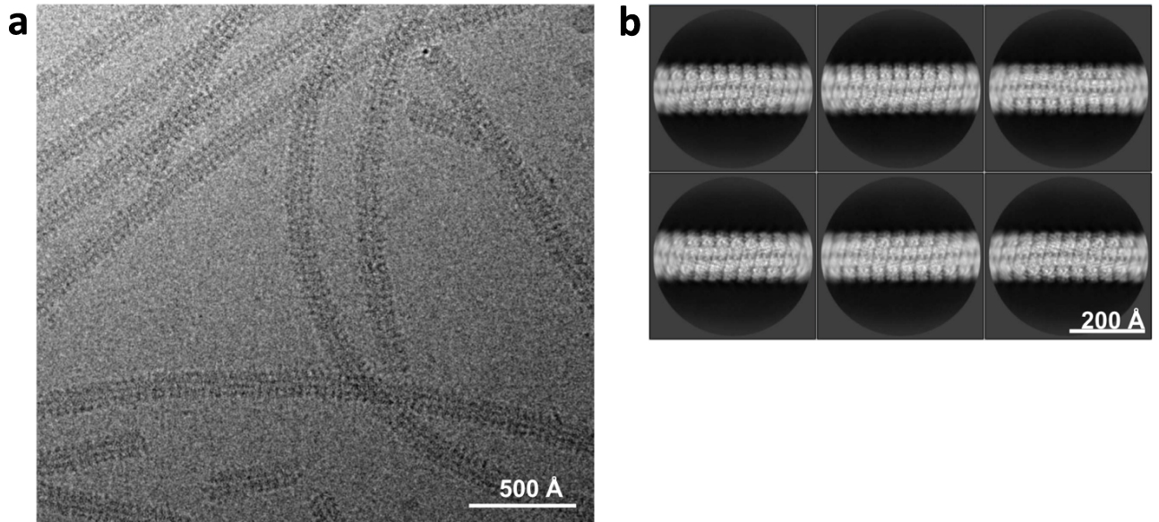
PVX particles contained on the grids were imaged at 300 keV with a K2 direct electron camera in the counting mode at 0.827 Å per pixel. From a total of 3029 movies collected, 2734 micrographs were selected for further analysis (Figure 1).

Figure 1: a) Representative area from a cryo-EM micrograph of a typical preparation of PVX virus showing its flexibility. b) Selection of typical 2D class averages of PVX filament used for helical reconstruction. Credit: A. Grinzato.
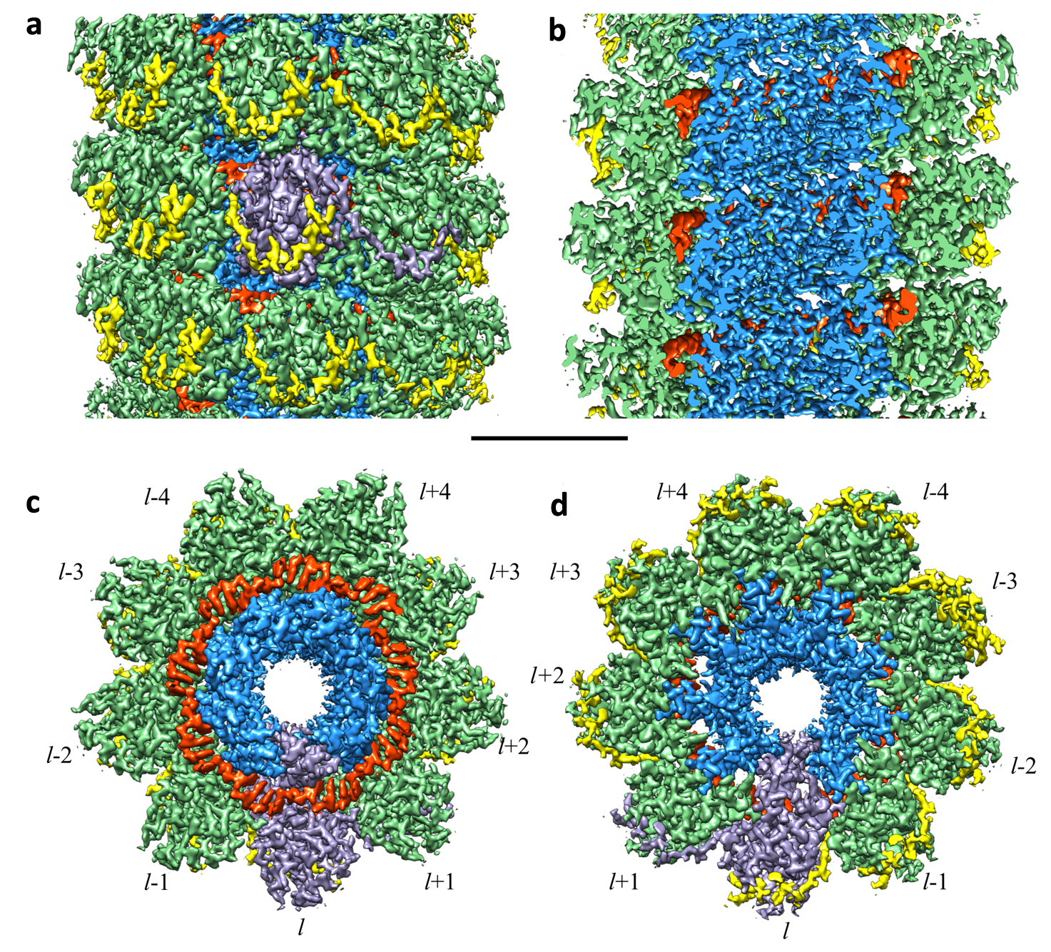
The reconstructions show that the virus is around 500 nm-long with a 15-nm diameter and is formed by repeat segments of coat proteins (CPs) arranged in a left-handed helical structure (Figure 2). The viral RNA is inserted in an internal crevice running the length of the particle, like a helter-skelter.

Figure 2: Cryo-EM structure of PVX virus at near-atomic resolution (CP N-terminal domain: yellow; CP core domain: green; CP C-terminal domain: blue; RNA: red). a) and b) Surface representation of the cryo-EM 3D reconstruction: (a) longitudinal front view, (b) cutaway view. c) and d) Close-up view of a section of nine consecutive CP protomers seen from (c) top and (d) bottom views. In each of the figures, where it is visible, a single CP is highlighted in purple. Scale bar: 30 Å. Credit: A. Grinzato.
The near-atomic resolution of the reconstruction means that both the virus’s structure and the protein–protein and protein–RNA interactions that are essential to its stability and function can be, for the first time, unambiguously described. Moreover, the high-resolution nature of the detail unveiled provides a platform for the structure-based design of antiviral compounds that are able to interfere with these interactions and to inhibit virus assembly.
Potato virus X can also be engineered to carry foreign RNA sequences or other chemicals and to introduce these into organisms.
“With our data, it is now possible to identify new sites for chemical and/or genetic modification of the virus,” explains Alessandro Grinzato of the Department of Biomedical Sciences at the University of Padua in Italy, and lead author of the paper.
“This could enable it to be used for a wide variety of nanotechnology applications, such as molecular imaging, drug delivery, vaccination, biosensor design, biomaterials development, and biocatalysis.”
Reference: Atomic structure of potato virus X, the prototype of the Alphaflexiviridae virus family, A. Grinzato et al., Nature Chem. Biol. (2020); doi: 10.1038/s41589-020-0502-4.
Text: Anya Joly.
Top image: See Figure 2.


