- Home
- Users & Science
- Scientific Documentation
- ESRF Highlights
- ESRF Highlights 2001
- Methods and Instrumentation
- Coherent Diffraction by Slits
Coherent Diffraction by Slits
High-brilliance third-generation synchrotron sources provide intense beams of sizes down to less than 10 µm. High angular resolution and small beam sizes need to be combined when micrometre scale objects are probed, as well as in ultra SAXS or in coherent scattering experiments. The beam size x and the resolution
q are, however, restricted by the diffraction limit:
x
q > 1/2. If slits are to be used to reduce the beamsize, it is a challenging task to obtain well-defined beams for small apertures, since in addition to strong diffraction phenomena, slits may cause intense parasitic streaks when the aperture has the same size as the typical length-scale of the surface roughness of the slit blades.
We have developed slits using polished cylinders to reduce the roughness. The performance of such a slit system depends on the radius and the material of the cylinders: either the incident photons are totally reflected from the cylinder of radius R (for incident angles i <
c) or they are absorbed by them (for
i >
c), provided that the penetration length µ-1 is small enough (µ-1 << 2R
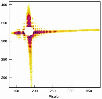
c). At 8 keV radiation, this is readily accomplished by molybdenum cylinders with a radius R = 1 mm [1]. A typical result obtained on ID1 in conditions of coherence is shown in Figure 158 with a 2 µm x 2 µm aperture. The diffraction pattern shows Fraunhofer interference fringes with a high contrast. Cross-terms of interference are also visible. The asymmetry of the diffraction pattern is explained by the intrinsic asymmetry of the slit, which can be determined quantitatively by analytical calculations [1,2].
 |
Fig. 158: Diffraction pattern of two crossed slits made of polished molybdenum cylinders. |
In Figure 158, a strong background is observed due to slit diffraction. For SAXS experiments this strong scattering needs to be further reduced by inserting a "guard slit" between pinhole and sample, the configuration of which is obviously dictated by two limiting cases: while closing the guard slits to the size of the pinhole aperture, they will develop their own diffraction pattern and opening them can only be done to an extent where they still reduce the diffraction of the aperture pinhole significantly. Moreover, the relative position of pinhole, guard slits and sample needs to be optimised. A too small distance between pinhole and guard slits is inefficient to reduce the background to signal ratio. On the other hand, it cannot be too large because of the front wave propagation. It can be shown [1] that for high resolution experiments, the guard slit has to be located close to the sample and at = a2/2*
from the pinhole (a is the aperture).
 |
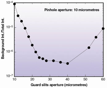
Fig. 159: Calculated background for a 10 µm pinhole for various apertures of the guard slit located 40 cm downstream. A clear background minimum appears for a 27 µm optimal aperture. |
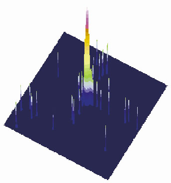
From diffraction theory one can estimate that the diffracted intensity of two successive slits, though strongly reduced, still decays with a q-2 dependence perpendicular to the beam. For a more precise calculation, numerical simulations have been performed [1], which result in a minimum of the parasitically diffracted intensity as a function of the slit opening (Figure 159). For a 10 µm pinhole at 8 keV for example, the slit diffraction can be reduced by more than two orders of magnitude for the guard slit closed to 27 µm and located a2/2* = 40 cm downstream. The experimental test of this prediction is displayed in Figure 160. It confirms that a well-chosen setting of two cylindrical slits permits the preparation of a well-defined primary X-ray beam with a peak to background ratio larger than 2 x 10⁴.
 |
Fig. 160: Primary beam intensity on a log scale obtained by using the experimental setup described in the text. The direct illumination CCD camera (pixel size: 22 µm) is located at 1.75 m from the guard slit. The primary beam has a maximum intensity of 2 x 10⁴ cps and covers 3 pixels only. |
References
[1] D. le Bolloc'h, F. Livet et al., Submitted to J. Synchrotron Rad.
[2] E. Vieg, S.A. De Vries, J. Alvarez and S. Ferrer, J. Synchrotron Rad. 4 210 (1997).
Authors
D. Le Bolloc'h (a,b), F. Livet (c), T. Schulli (a), F. Bley (c), H.T. Metzger (a) and M. Veron (c)
(a) ESRF
(b) Present address: Laboratoire de Physique des Solides, Orsay (France)
(c) LTPCM-ENSEEG, UMR-CNRS/INPG/UJF BP75, Saint Martin d'Hères (France)


