Other Materials
Other more "exotic" materials have been also studied at ID21, such as patina from Dogon statues, varnish from musical instruments or colour photographs as well.
|
M. Cotte, T. Fabris, J. Langlois, L. Bellot‐Gurlet, F. Ploye, N. Coural, C. Boust, J. P. Gandolfo, T. Galifot and J. Susini, "Rediscovering Ducos du Hauron's Color Photography through a Review of His Three‐Color Printing Processes and Synchrotron Microanalysis of His Prints", Angewandte Chemie, 57, 7364-7368 (2018). J. Langlois, F. Ploye, M. Cotte, K. Théard, L. Clivet, E. Laval, P. Bydlowski, J. P. Gandolfo, C. Boust and N. Coural, "Characterization of Louis Ducos du Hauron’s diaphanies", ICOM-CC 18th Triennial Conference, Copenhagen, (2017). |
Louis Ducos du Huron (1837–1920) dedicated his entire life to the elaboration of physical–chemical processes for color photography. This study aimed at highlighting his unique contribution to three‐color printing through 1) an in‐depth review of the many protocols he published and 2) the synchrotron‐based IR and X‐ray microanalysis of fragments sampled in three artworks. Ducos du Hauron's method relied on the preparation and assembly of three monochromes (red, blue, yellow). This study brings to light complex multistep recipes based on photochemistry (carbon print), organic, and inorganic chemistry. The various ingredients involved (e.g., pigments, dichromate gelatin, collodion, resin) were identified and localized through their spectroscopic signature, confirming the relevance of synchrotron spectromicroscopy for the characterization of historical photographs. The impressive correlation between texts and chemical analyses calls for a wider application to the history of photography.
|
V. Mazel, P. Richardin, D. Debois, D. Touboul, M. Cotte, A. Brunelle, P. Walter and O. Laprévote, "Identification of ritual blood in African artifacts using TOF-SIMS and synchrotron radiation microspectroscopies", Analytical Chemistry, 79, 9253-9260 (2007). |
A new protocol is implemented to demonstrate the presence of blood in the patina of African art objects from Mali. Divided into three steps, the protocol first consists in demonstrating the presence of proteins and localizing them in the sample's cross sections using time-of-flight secondary ion mass spectrometry (TOF-SIMS) and synchrotron-based infrared microspectrometry (μFT-IR). In a second time, TOF-SIMS is used to investigate heme, which is a blood marker. If heme is missing, which could mean that it is too degraded to be detected, X-ray microfluorescence (μXRF) and X-ray absorption near-edge microspectroscopy (μXANES) are used to prove the presence of iron in the protein area and to get a fingerprint of its chemical environment. This permits us thus to demonstrate that iron is indeed linked with proteins and not with mineral phases of the sample. Coupled with the ritual context of the objects, this constitutes a proof of the use of blood. Thanks to this protocol, which has the major advantage of avoiding false positive results, the presence of blood has been demonstrated in seven out of the eight studied samples.
 |
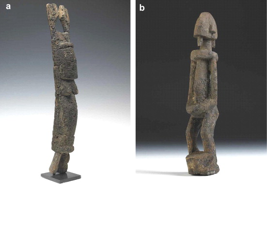
V. Mazel, P. Richardin, D. Debois, D. Touboul, M. Cotte, A. Brunelle, P. Walter and O. Laprévote, "The patinas of the Dogon-Tellem statuary: A new vision through physico-chemical analyses", Journal of Cultural Heritage, 9, 347-353 (2008). |
Numerous African art objects collected in the course of colonial or ethnological expeditions during the 20th century are partially or completely covered with a so-called ‘‘patina’’. These patinas have been formed during religious and ritual ceremonies, where different substances have been spread out at the surface of the objects. The anthropomorphic statuettes from the Dogon culture are well-known examples of this kind of practice. A better understanding of the chemical composition of these patinas could explain details of the ceremonial practices realized throughout the centuries. An important challenge is to extract unequivocal information about the original constituents of these patinas, and the techniques used for their application on ritual objects. The Dogon statuary can be divided into three periods. The oldest one, before 1350 A.D., is called the Tellem period, according to the name of the people who lived in this area before the arrival of the Dogon. The most recent, after 1650 A.D., is the Dogon period. These two periods are separated by an intermediary period, where the attribution to the Tellem or to the Dogon cultures is difficult. During our study, we have worked on the patina of 12 Dogon artifacts from these three different periods. In this paper, we present the results obtained for two representative objects, one of the Tellem period and one of the Dogon period. The aim is to access the chemical composition of the patinas and to see if they can be compared to ethnological reports, as is often done in the literature on ethnological studies. Chemical imaging techniques, based on the combination of microscopy and spectroscopy, are well suited to study both the composition and the spatial organization of heterogeneous complex mixtures of organic and inorganic matter. Time-of-flight secondary ion mass spectrometry (ToF-SIMS), followed by scanning electron microscopy with energy dispersive X-ray analysis (SEM-EDS), and synchrotron radiation-based Fourier transform infrared microscopy (SR-µFTIR) have been applied to non-destructive analyses of micro-samples of the patinas of several Dogon statuettes. A very careful preparation, using ultramicrotomy on embedded samples, allowed us to perform successively all these measurements on a single fragment. Comparison and superposition of the different chemical images lead us to identify minerals (clays, quartz and calcium carbonate), and different organic products (proteins, starch, lipids), as well as to map their spatial distribution. The patina of the most recent statuettes presents a stratigraphy which can be related to successive uses of the objects for ritual purposes, and the results are in agreement with the ethnological reports. For the Tellem objects, the significant quantity of minerals and the lack of stratigraphy suggest that the patina was deposited according to a different recipe. This difference suggests the existence of a diversity of rituals that are more extensive than that described by the ethnologists.
|
J. P. Echard, M. Cotte, E. Dooryhée and L. Bertrand, "Insights into the varnishes of historical musical instruments using synchrotron micro-analytical methods", Applied Physics A, 92, 77-81 (2008). |
Though ancient violins and other stringed instruments are often revered for the beauty of their varnishes, the varnishing techniques are not much known. In particular, very few detailed varnish analyses have been published so far. Since 2002, a research program at the Musée de la musique (Paris) is dedicated to a detailed description of varnishes on famous ancient musical instruments using a series of novel analytical methods. For the first time, results are presented on the study of the varnish from a late 16th century Venetian lute, using synchrotron micro-analytical methods. Identification of both organic and inorganic compounds distributed within the individual layers of a varnish microsample has been performed using spatially resolved synchrotron Fourier transform infrared microscopy. The univocal identification of the mineral phases is obtained through synchrotron powder X-ray diffraction. The materials identified may be of utmost importance to understand the varnishing process and its similarities with some painting techniques. In particular, the proteinaceous binding medium and the calcium sulfate components (bassanite and anhydrite) that have been identified in the lower layers of the varnish microsample could be related, to a certain extent, to the ground materials of earlier Italian paintings.


