- Home
- News
- General News
- #EBSstories Tackling...
#EBSstories Tackling a deadly cardiovascular disease
06-05-2021
A deadly condition that starts as a tear in the aorta has been reproduced on ID17 by scientists from the École de Mines de Saint Etienne in France, with the aim of better understanding the onset of the disease.
Aortic dissection is a life-threating disease that occurs suddenly when a tear appears in the aorta and this tear allows the blood flow between the layers of the aortic wall. The disease’s onset can take place anywhere in the aorta, but it is often near the heart. Although it is rare, affecting three every 100000 people, its mortality rate is high and, if untreated, 50% of people die within 24 hours.
“The mechanisms underlying this disease are not really well understood, mainly due to the fact that aortic dissection is very difficult to observe, because it is internal and it is a very rapid process”, explains Joseph Brunet, scientist in bioengineering at the École de Mines de Saint Etienne.
The ESRF, with its new EBS, provided the perfect stage for the scientists to re-enact the disease in an aorta. “The aorta is extremely heterogeneous and it is composed of many microstructural components. Only with EBS we are able to observe these microelements and the global structure of the aorta at the same time, with amazing resolution in time and in space, on beamline ID17”, explains Brunet.
 |
 |
|
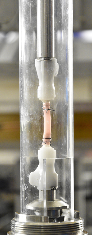
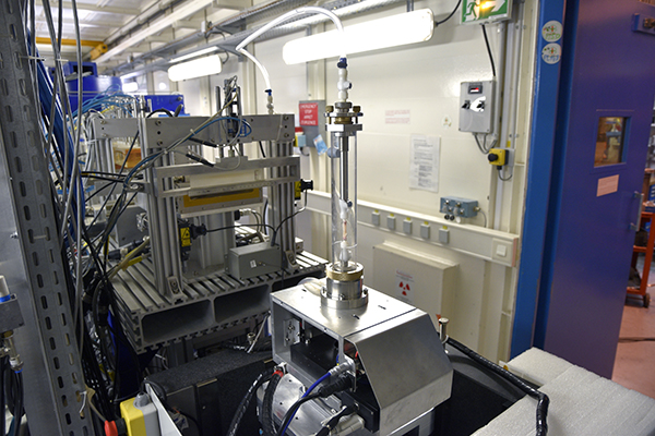
On the left, an aorta section mounted on the sample holder. On the right, the set up on ID17. Credits: J. Brunet. |
|
The team studied how this tear propagates. Previously, the scientists designed a custom-made tension-inflation device suited for X-ray microtomography to reproduce the movements that take place in the aorta when breathing. This device, combined with X-ray tomography on ID17, allowed them to follow the dissection propagation in vitro in its early stages. Results will provide them with precise information on the arterial dissection mechanics, in particular, critical pressure, direction of the propagation, preferred path within the layered microstructure, strain field around the tear and also possible involvement of different fracture modes.
“It is the first time that aorta dissection is studied in situ using synchrotron radiation, to my knowledge, and we are very excited because we expect that it will shed light on the mechanical phenomena leading to arterial dissection”, he concludes.
Text by Montserrat Capellas Espuny. Video by Montserrat Capellas Espuny and Mark McGee.
.
partners


European Synchrotron Radiation Facility - 71, avenue des Martyrs, CS 40220, 38043 Grenoble Cedex 9, France.