- Home
- News
- General News
- How do asbestos...
How do asbestos fibres lead to cancer? #weekendusers
24-11-2017
Asbestos has been forbidden for some years in many countries around the world. Yet the peak of mortality related to asbestos-related diseases will take place in the next decade, as it takes a long time to develop the disease. Researchers from the Italian national research council (CNR-Nanotec) (Rome, Italy) hope that by studying the interactions between asbestos with host organisms can lead to develop more efficient medical treatments or prevention policies. They are on ID16A looking at the nanoscale at the elements making up the coating developing around asbestos once in the lungs.
“I started with this project when I was living in Torino, where two of the biggest factories of asbestos in Europe were based in the past. I met a professor who was studying the subject and I thought it was fascinating”, explains Fabrizio Bardelli, researcher at CNR-Nanotec. “We all think that asbestos is something from the past, but we still suffer the consequences today”, he adds.
Indeed, asbestos fibres can enter in living organisms by inhalation and, due to its high bio-persistence, can manifest its toxicity after long (20 to 40 years). For these reasons, since the 1990s asbestos started to be banned in many countries and, with a few relevant exceptions (Russia, China, Canada, and Brazil), it is almost abolished today.
 |
 |
|
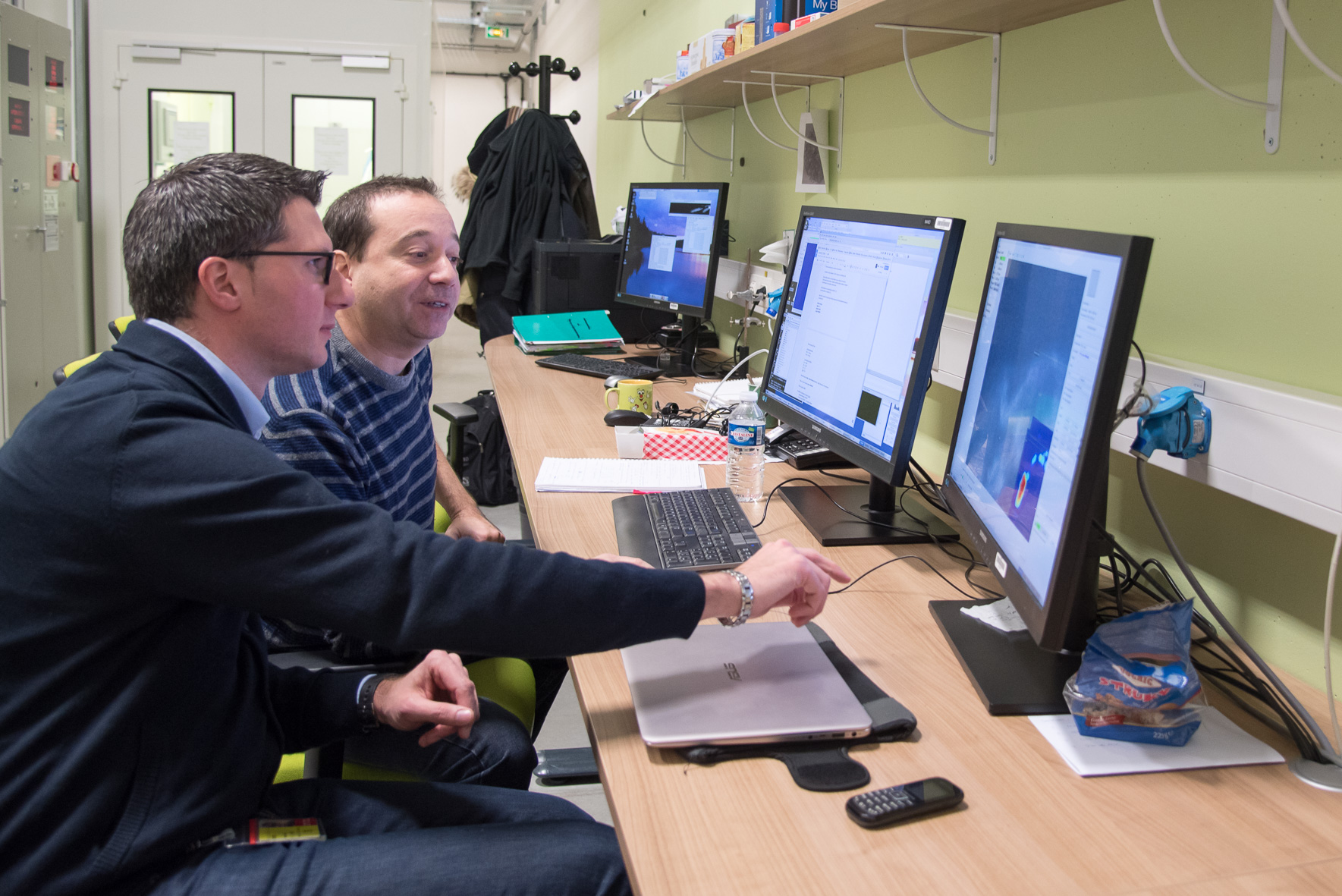
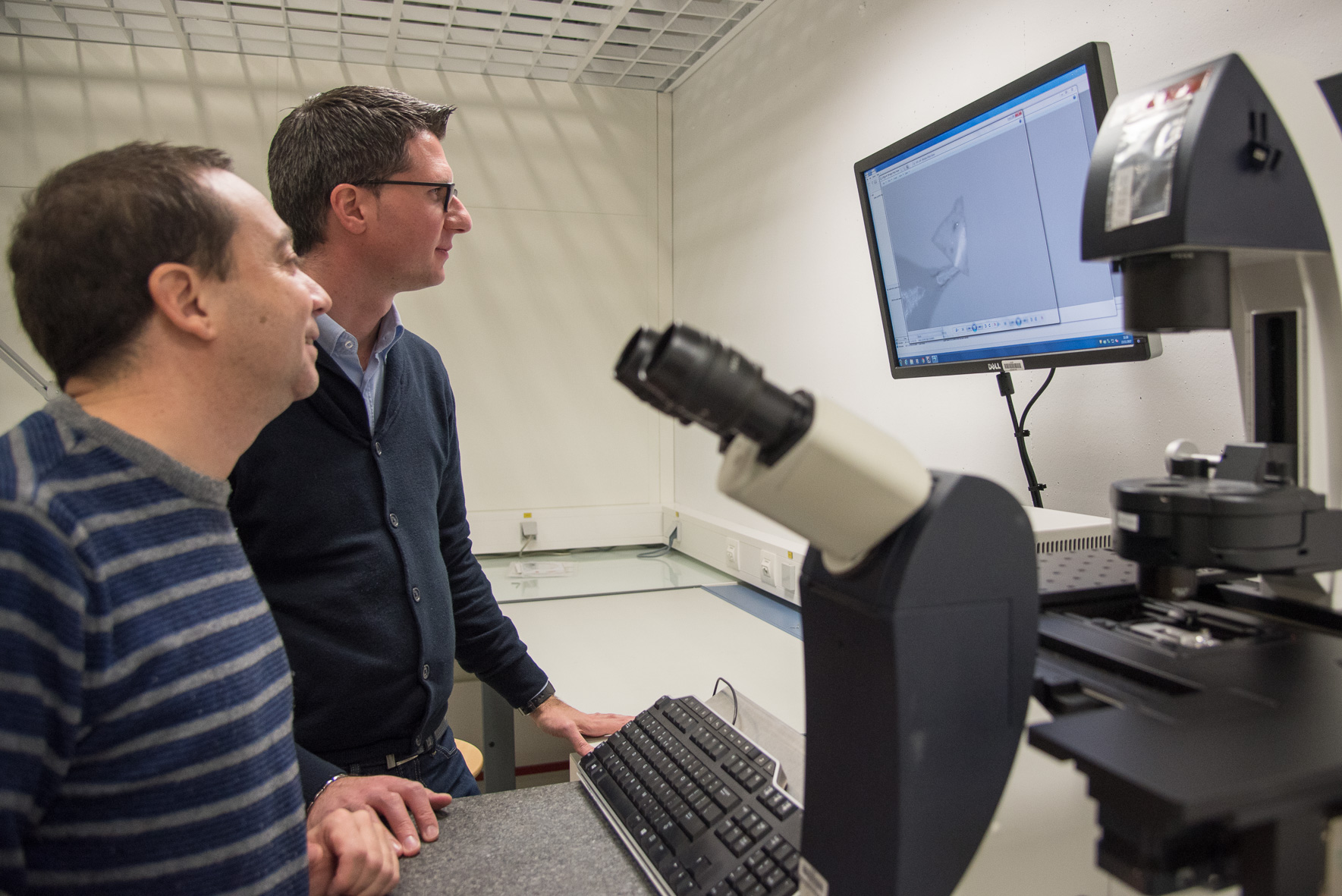
Fabrizio Bardelli (with stripy top) and Francesco Brun on ID16A and in the lab. Credits: C. Argoud. |
|
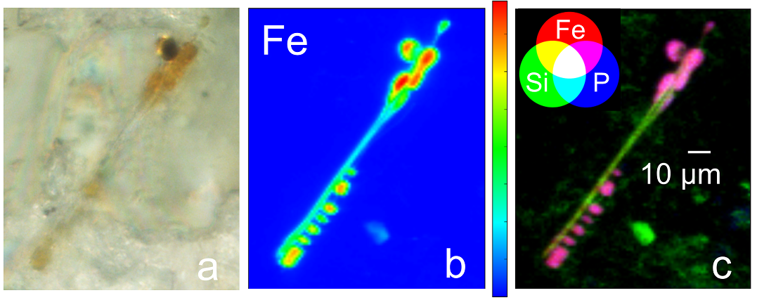
Exposure and contamination to asbestos can originate from several diverse sources, such as asbestos products plants, steel plants, shipyards, and asbestos mines. In the lungs, iron-containing asbestos fibres irritate the tissue, causing minerals and proteins to cluster around the foreign fibres, in a process known as biomineralization. The resulting clusters are known as ‘asbestos bodies’ and are typical of mesothelioma, a deadly cancer of the lung lining, and of other asbestos-diseases, such as asbestosis.
Bardelli and his colleague, researcher Francesco Brun are now spending a week testing samples of lung tissue affected by asbestos. It is the second part of an experiment that started last year on ID16A: “The first experiment went very well, but it was only a small number of samples. We want to increase the samples so that we can compare them all and have better statistics”, explains Brun. During this beamtime they will be running 24h long scans, so no sleepless nights for them. “The tricky part of this experiment is the preparation of the sample and the mounting, once it is done it isquite straight forward”, he adds.
 |
|
(a) Optical microscope image (500x) of an asbestos body. (b) Elemental distribution map of Fe of the same asbestos body (the color bar indicates the concentration of Fe), done in a previous experiment on ID21. (c) RGB color combination showing the distribution and colocalization of Si, Fe, and P. |
Extensive research on asbestos interaction with host organism is essential to better understand the pathogenesis, and, in this way, to develop more efficient medical treatments or prevention strategies. Bardelli says that the benefits of their research go beyond this: “our research on the mechanisms inducing the chain of events leading to mesothelioma can help to foresee the toxicity of new man-made nanofibers, such as metal-oxide or carbon nanotubes, which are becoming of more and more widespread use in electronic devices”.
This research project received European funds through a Marie Sklodowska-Curie Individual Fellowship action (BiominAB-3D, H2020 - GA707905; http://biominab3d.altervista.org)
Text by Montserrat Capellas Espuny
Top image: Asbestos roofing sheets. Credits: Nick.
partners


European Synchrotron Radiation Facility - 71, avenue des Martyrs, CS 40220, 38043 Grenoble Cedex 9, France.