- Home
- News
- General News
- Towards an improved...
Towards an improved treatment for tetanus infection
18-10-2021
Scientists led by the University of Padova have found a new candidate treatment against tetanus neurotoxins with advantages over the one currently used. The cryo-electron microscope of the ESRF has assisted them to elucidate its mechanism. The results have been published in the Journal of Clinical Investigation this week.
Tetanus bacteria are commonly found in soil and the manure of animals such as horses and cows. If the bacteria enter the body through a wound, they can quickly multiply and release a potent toxin that affects the nerves, causing symptoms such as muscle stiffness and spastic paralysis. About 10% of cases prove to be fatal.
A vast proportion of the population is vaccinated against the tetanus neurotoxin produced by this bacterium, but when someone turns up in a hospital with a potentially infected wound, doctors immediately apply anti-tetanus immunoglobulin. This cure contains antibodies, made from either human or horse blood plasma, to prevent the tetanus neurotoxin from working. First authors Marco Pirazzini from University of Padova explained the limitations of the current treatment: “It is made up of a mix of antibodies, and only a minor fraction of them can neutralize the toxin. Moreover, it is contained in a serum, which entails the same risks as a blood transfusion”.
Now a team of scientists of the University of Padova (Italy), Università della Svizzera Italiana (Switzerland), Humabs BioMed SA (Switzerland), University College London (UK), Istituto Nazionale Genetica Molecolare (Italy) and the ESRF has found an alternative to the current prophylaxis for tetanus infection. It consists of two monoclonal antibodies that block the toxin from entering in the cell. “We screened lots of antibodies, discarded the worst while choosing the two best neutralizing ones, which were produced recombinantly. So we need less quantity of antibodies and the targeted treatment is equally effective but without the risks associated with the serum”, Pirazzini said.
 |
|
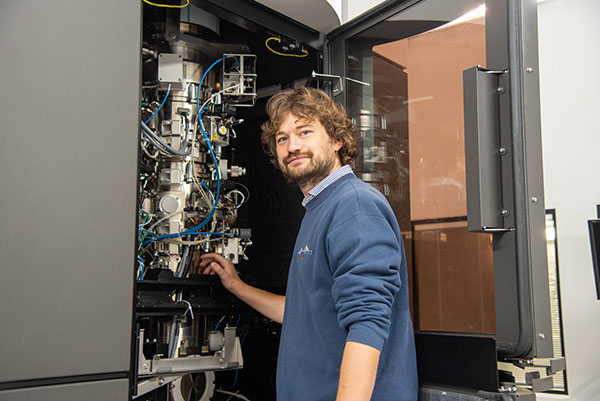
Alessandro Grinzato using the Cryo-electron microscope at the ESRF. Credits: C. Argoud. |
Pirazzini and colleagues had to go a long way before reaching these conclusions. First, they tried to crystallize the antibody protein interacting with the toxin, but it did not work. So then, they decided to move to cryo-electron microscopy (cryo-EM) at the ESRF to try to find some answers, as it doesn’t require crystals, but it wasn’t easy. “The structures were constantly moving, as they are very flexible, which made for an extremely challenging experiment. But we persevered and ended up solving the structure and describing the flexibility of the different domains”, explained first author Alessandro Grinzato, previously at the University of Padova and now a post-doctoral researcher at the ESRF.
The constant movement allowed the scientists to identify which parts of the toxin are bound by the two antibodies. They found that one antibody binds the domain used by the toxin to get anchored to the neuron surface, sticking exactly in between two parts used by the toxin to interact with its two receptors. This antibody prevents the toxin binding to neurons. The second antibody interferes with a subsequent step of the process of toxin entry inside neurons as it then freezes the structural change necessary to deliver the catalytic domain, i.e. the part of the toxin responsible for the neuroparalytic activity, into the neuron cytoplasm.
The next step for the team is to start clinical trials: “We are very optimistic about this medicine and hope that it can receive approval for use in the coming years as safe therapeutics for the prophylaxis and therapy of tetanus”, concluded Grinzato.
Reference:
Pirazzini, M. et al, J Clin Invest. 2021. https://doi.org/10.1172/JCI151676.
Text by Montserrat Capellas Espuny.
partners


European Synchrotron Radiation Facility - 71, avenue des Martyrs, CS 40220, 38043 Grenoble Cedex 9, France.