- Home
- News
- Spotlight on Science
- A new charge reordering...
A new charge reordering in magnetite
14-12-2021
A new charge reordering in magnetite was discovered at beamline ID26 using core-level X-ray spectroscopy. This reordering takes place in three steps and maps the temperature-dependence of the electrical conductivity and magnetism up to the Curie-temperature.
Magnetite (Fe3O4) is one of the most abundant Fe-bearing minerals on Earth, and it finds many applications in areas such as paleomagnetism, medicine, data recording and engineering. It is a prototypical magnetic material and was used for the first compass. Understanding the ordering of the charges in magnetite is one of the oldest questions in solid state physics, as magnetite exhibits a first-order metal-to-insulator transition at TV~123 K [1]. Fe ions reside in two interstitial sites in Fe3O4: (i) Fe2+ and Fe3+ in octahedral sites; and (ii) Fe3+ in tetrahedral sites. Several decades of experimental and theoretical research have revealed that an intricate charge and orbital ordering takes place below the metal-to-insulator transition temperature, where the Fe ions at the octahedral sites form a network of Fe3+-Fe2+-Fe3+ linear molecules (called trimerons) [2]. Short-range structural fluctuations linked to the presence of trimerons have been recently shown to exist in the high-temperature metallic-like phase [3]. However, the role of these short-range trimerons and the mechanism behind their collapse remains an unsolved puzzle. This motivated the design of a new experiment that addresses these questions.
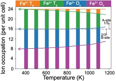
Fig. 1: Occupation of Fe3+ and Fe2+ ions in octahedral (Oh, plotted in purple and blue) and in tetrahedral (Td, plotted in green and orange) sites per unit cell of Fe3O4 shown for the temperatures between 300 - 1200 K, where charge redistribution is observed. The vertical bars are shown to guide the eye. Note that the total Fe charge remains constant.
Fluorescence-detected X-ray absorption spectroscopy with high energy resolution was used at beamline ID26 to investigate the short-range structural fluctuations and charge ordering in magnetite. In this experiment, a 1s core-electron was resonantly excited to the empty 3d orbitals of Fe, providing an element- and site-selective probe of the Fe ions in Fe3O4. A new charge reordering was found to occur in the metallic-like phase, where the extra electron of the Fe2+ ion at the octahedral sites is transferred progressively to the tetrahedral sites with increasing temperature (Figure 1). This implies that the octahedral sublattice becomes hole-doped and, hence, the Fe3+-Fe2+-Fe3+ trimerons are altered. It was found that the level of hole-doping can be continuously (and reversibly) tuned by controlling the temperature, and three regimes of self-doping were identified that describe the temperature-dependence of key physical properties such as the electrical conductivity and magnetism.
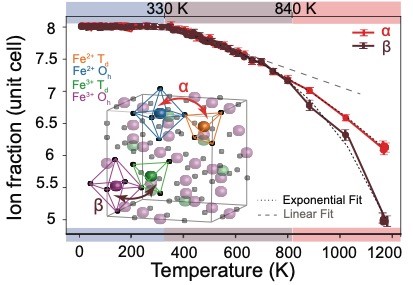
Fig. 2: Temperature evolution of the redistribution parameters α (red) and β (brown) in Fe3O4. Linear and exponential fits are plotted in dashed grey and black dashed lines. A cartoon of the ion transfer in a unit cell of Fe3O4 is shown in the inset. The colour code on the x axis of the figure illustrates the regimes described in the text.
Figure 2 shows the temperature-induced hole-doping described by two parameters, α and β, according to the formula: α Fe2+Oh +β Fe3+Td + (8 - α) Fe2+Td + (16 - β) Fe3+Oh. The first regime is identified by α = β = 8, meaning that no charge transfer effect takes place (highlighted in blue in Figure 2). Here, short-range Fe3+-Fe2+-Fe3+ trimerons exist as sketched in the left of the featured figure. The second regime is identified by α = β < 8, where the extra electron at the octahedral site is transferred to the tetrahedral sites (highlighted in brown in Figure 2). In this regime, the self-doping modifies the trimerons and leads to their gradual destruction (middle sketch of the featured figure). The onset of the third regime is identified by α ≠ β (highlighted in red in Figure 2). This implies that cations (and not only charge) are exchanged between the two sublattices where the occupation of the interstitial B sites increases. The trimerons do not exist in this regime (last sketch of the featured figure).
These results shed light on the role of trimerons: short-range trimerons act as descriptors of correlations (i.e., they describe the evolution of electrical and magnetic properties) in magnetite, and they collapse due to the temperature-driven hole self-doping. The results provide an elegant analogy between the effect of chemical doping (where the ratio of Fe2+/Fe3+ ions is chemically modified by adding impurity atoms of different valency) and temperature-driven self-doping in magnetite, and inspires the question: can this analogy be generalised to other systems exhibiting the metal-insulator transition? The hard X-ray probe at beamline ID26 makes it possible to explore a large range of experimental parameters, and future research will include studying magnetite under high pressure and high temperature.
Principal publication and authors
Temperature-Driven Self-Doping in Magnetite, H. Elnaggar (a,b), S. Graas (a), S. Lafuerza (c), B. Detlefs (c) W. Tabiś (d,e), M. Gala (d), A. Ismail (a), A. van der Eerden (a), M. Sikora (f), J. Honig (g), P. Glatzel (c), F. de Groot (a), Phys. Rev. Lett. 127, 186402 (2021); https://doi.org/10.1103/PhysRevLett.127.186402
(a) Debye Institute for Nanomaterials Science, Utrecht (The Netherlands)
(b) Sorbonne Université, CNRS UMR 7590, Institut de Minéralogie, de Physique des Matériaux et de Cosmochimi, Paris (France)
(c) ESRF
(d) Faculty of Physics and Applied Computer Science, AGH University of Science and Technology, Krakow (Poland)
(e) Institute of Solid State Physics, TU Wien, Vienna (Austria)
(f) Academic Centre for Materials and Nanotechnology, AGH University of Science and Technology, Krakow (Poland)
(g) Department of Chemistry, Purdue University, West Lafayette, Indiana (USA)
References
[1] E.J.W. Verwey, Nature 144, 327 (1939).
[2] M.S. Senn et al., Nature 481, 173 (2012).
[3] G. Perversi et al., Nat. Commun. 10, 2857 (2019).
partners


European Synchrotron Radiation Facility - 71, avenue des Martyrs, CS 40220, 38043 Grenoble Cedex 9, France.