- Home
- News
- Spotlight on Science
- A “spooky” approach...
A “spooky” approach towards high-fidelity X-ray imaging for all
24-02-2022
High-fidelity X-ray imaging frequently requires elaborate detection schemes. Hard X-ray ghost imaging, on the contrary, allows one to deploy a rudimentary detection approach: a ‘bucket’ detector with no pixels. Previously, this reduced image quality, a challenge now overcome at the ESRF.
The value of X-ray imaging as a medical tool has been known worldwide since Röntgen’s famous first radiograph showed the bones in the hand of his wife, Anna Bertha Ludwig. One important aspect of this success story is the robustness of the method. X-rays do not only transmit through samples, but they face basically no scattering on the length scales relevant for radiography. Only a source and a photographic film, but no other optical element, are required to produce ‘sharp’ pictures.
A century later, research is focused on advancing X-ray imaging by increasing the contrast exploiting the full complex-valued refractive index of matter (frequently associated with X-ray phase contrast imaging), reaching for higher spatial resolution while maintaining an acceptable dose. The results achieved are impressive but frequently sacrifice one of the fundamental properties of X-ray imaging from the early days: the robustness mentioned above. New developments face challenges during their introduction in real-world applications if they require large-scale facilities such as synchrotron light sources or detection schemes, which only a few people in the world can operate.
Hard X-ray ghost imaging uses a radically different approach. Instead of complicated illumination schemes coupled to high-end detectors, it requires a defined structure in the X-ray optical beam path as well as a detector with no pixels (also called a ‘single-pixel camera’ or ‘bucket’ detector). The complexity of the acquisition is no longer the responsibility of the optical hardware and is shifted to computational algorithms, which reveal the final image out of a set of rather noisy patterns. Hence, the technique is frequently also considered as ‘computational’ X-ray ghost imaging because computational reconstruction is a ‘software lens’ that forms an intrinsic part of the imaging system. The proof-of-concept of hard X-ray ghost imaging was previously carried out at the ESRF [1,2] including a demonstration of ghost tomography [3] but, up to now, images suffered from limited contrast and spatial resolution.
An approach has now been developed at beamline ID19 that exploits structured-detection, single-pixel imaging to produce an X-ray ghost image with phase contrast, accuracy and high fidelity. As a gold standard reference, a high-fidelity, synchrotron-based, phase-contrast radiography of an aluminium sponge was recorded with a state-of-the art area detector (Figure 1a). The same sample was then scanned using the new ghost imaging protocol, with a rudimentary detector and an image calculated from the set of corresponding illumination patterns. The resulting X-ray phase-contrast ghost image (Figure 1b) provides accurate information regarding density variations in the sample and visibly renders edges that are otherwise invisible with X-ray attenuation contrast. This shows how the previously separate fields of X-ray phase contrast and X-ray ghost imaging can be merged.
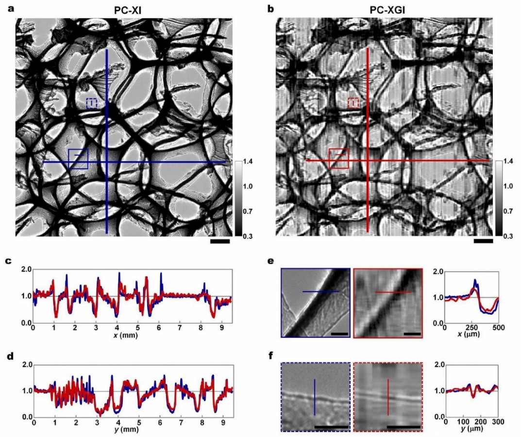
Fig. 1: X-ray transmission images of interconnected aluminium lamellae cut from a sponge sample. a) Phase-contrast X-ray image (PC-XI): radiographs directly recorded using a 2D imaging detector. b) Phase-contrast X-ray ghost image (PC-XGI): calculated from synthesised ghost images. Scale bars in (a) and (b), 1 mm. c) Horizontal line profiles. d) Vertical line profiles. e,f) Magnified views of the insets in (a) and (b) showing phase-contrast enhancement at the edges of representative thick and thin regions of the lamellae. Scale bars in (e) and (f), 250 µm.
This demonstration of phase-contrast ghost imaging with X-rays has the potential to improve X-ray ghost imaging from its current status as a niche technique to a routinely applied method. In future, it would allow the acquisition of high-fidelity X-ray images, for example, in harsh environments or under-developed countries, as it would only require a source, a set of X-ray masks and a semiconductor block USB-plugged to a smart phone to produce X-ray images.
Principal publication and authors
X-ray phase-contrast ghost imaging using a single-pixel camera, M.P. Olbinado (a,b), D.M. Paganin (c), Y. Cheng (a), A. Rack (a), Optica 8, 1538 (2021); https://doi.org/10.1364/OPTICA.437481
(a) ESRF
(b) Paul Scherrer Institut, Villigen (Switzerland)
(b) School of Physics and Astronomy, Clayton (Australia)
References
[1] D. Pelliccia et al., Phys. Rev. Lett. 117, 113902 (2016).
[2] D. Pellicia et al., IUCrJ 5, 428-438 (2018).
[3] A. Kingston et al., Optica 5, 1516 (2018).
partners


European Synchrotron Radiation Facility - 71, avenue des Martyrs, CS 40220, 38043 Grenoble Cedex 9, France.