- Home
- News
- General News
- Scientists discover...
Scientists discover 356 animal inclusions trapped in 100 million years old opaque amber
31-03-2008
Paleontologists from the University of Rennes (France) and the ESRF have found the presence of 356 animal inclusions in completely opaque amber from mid-Cretaceous sites of Charentes (France). The team used the X-rays of the European light source to image two kilogrammes of the fossil tree resin with a technique that allows rapid survey of large amounts of opaque amber. At present this is the only way to discover inclusions in fully opaque amber.
Opaque amber has always been a challenge for paleontologists. Researchers cannot study it because the naked eye cannot visualize the presence of any fossil inclusion inside. In the Cretaceous sites like those in Charentes, there is up to 80% of opaque amber. It is like trying to find, in complete blindness, something that may or may not be there.
However, the paleontologists Malvina Lak, her colleagues from the University of Rennes and the ESRF paleontologist Paul Tafforeau, together with the National Museum of Natural History of Paris, have applied to opaque amber a synchrotron X-ray imaging technique known as propagation phase contrast microradiography. It sheds light on the interior of this dark amber, which resembles a stone to the human eye. “Researchers have tried to study this kind of amber for many years with little or no success. This is the first time that we can actually discover and study the fossils it contains”, says Paul Tafforeau.
 |
|
|
Figure 1: Pieces of opaque amber. Image credits: V. Girard/D. Néraudeau, UMR CNRS 6118. |
The scientists imaged 640 pieces of amber from the Charentes region in southwestern France. They discovered 356 fossil animals, going from wasps and flies, to ants or even spiders and acarians. The team was able to identify the family of 53% of the inclusions.
Most of the organisms discovered are tiny. For example, one of the discovered acarians measures 0.8 mm and a fossil wasp is only 4 mm. “The small size of the organisms is probably due to the fact that bigger animals would be able to escape from the resin before getting stuck, whereas little ones would be captured more easily”, explains Malvina Lak.
|
|
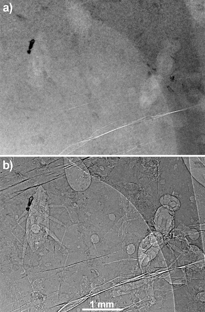
|
Figure 2: a) radiography of an amber block with inclusions in absorption mode. b) the same radiograph in propagation phase contrast mode with 990 mm of propagation distance (pixel size: 5 µm). Credits: M. Lak, P. Tafforeau, D. Néraudeau (ESRF Grenoble and UMR CNRS 6118 Rennes). |
Water to see tiny fossils better
The surface features of amber pieces, like cracks, stand out more in the images than the fossil organisms in the interior when using synchrotron radiation. In order to solve this problem, scientists soaked the amber pieces in water before the experiment. Because water and amber have very similar densities, immersion made the outlines of the amber pieces and the cracks almost invisible. At the same time, it increased overall inclusion visibility, leading to better detection and characterization of the fossils.
Classification of species
Once discovered on the radiographs, some of the organisms were imaged in three dimensions and virtually extracted from the resin. The high quality of these 3D reconstructions enables paleontologists to precisely study and describe the organisms. The success of this experiment shows the high value of the ESRF for the study of fossils. “Opaque amber hosts many aspects of past life on our planet that are still unknown, and the use of third generation synchrotron sources will continue to play an important role in unveiling them”, asserts Malvina Lak.
|
|
|
Figure 3: Examples of virtual 3D extraction of organisms embedded in opaque amber: a) Gastropod Ellobiidae; b) Myriapod Polyxenidae; c) Arachnid; d) Conifer branch (Glenrosa); e) Isopod crustacean Ligia; f) Insect hymenopteran Falciformicidae. Credits: M. Lak, P. Tafforeau, D. Néraudeau (ESRF Grenoble and UMR CNRS 6118 Rennes). |
Reference
M. Lak, D. Néraudeau, A. Nel, P. Cloetens, V. Perrichot and P. Tafforeau, Phase Contrast X-ray Synchrotron Imaging: Opening Access to Fossil Inclusions in Opaque Amber, Microscopy and Microanalysis, Forthcoming article doi:10.1017/S1431927608080264.
Further Information
- Animation of 3D images obtained from the experiment: Amber_animation.mov (57 mb); Amber_animation.avi (38 mb)
partners


European Synchrotron Radiation Facility - 71, avenue des Martyrs, CS 40220, 38043 Grenoble Cedex 9, France.