- Home
- Users & Science
- Scientific Documentation
- ESRF Highlights
- ESRF Highlights 2006
- X-ray Imaging and Optics
- Holotomography reveals new features in seed structure
Holotomography reveals new features in seed structure
Seeds are central to the world’s wildlife as well as to its agriculture, in the latter case both in the form of cereals and as the mediator for the renewal of crops. Seeds of the botanical geneticist’s favourite test plant, the weed-like, unassuming Arabidopsis thaliana (wall cress or mouse-ear cress) were investigated using holotomography, an approach developed at the ESRF [1]. This technique is quantitative phase tomography with micrometre resolution using hard synchrotron radiation X-rays. It is a variant of microtomography, the microscopy-scale version of medical computed tomography (CT). Many images are taken for different orientations of a given sample and then digitally combined to provide either three-dimensional (3D) renditions or virtual slices along any direction. Tomography usually relies on attenuation or absorption contrast, i.e. to the local variations in its amplitude or intensity, with respect to the average beam intensity, due to the inhomogeneous distribution of absorption in the sample. For holotomography, an essential contribution to the image is due to the effect of the sample on the phase of the X-ray beam going through it. This is due to inhomogeneous refractive index and/or thickness distributions in the sample. At a third-generation synchrotron radiation source such as ESRF, these relative phase variations can be turned very simply into contrast and thus produce an image. Thanks to the spatial coherence in the beam, especially on a long beamline such as the Imaging Beamline ID19, Fresnel diffraction turns local variations in phase into changes in intensity, and thus into an image. This requires no lens, but the detector must be placed some distance from the sample. The images recorded at more than one distance must then be processed to provide quantitative phase maps. Phase maps corresponding to different sample orientations are then combined, using the same software as for absorption tomography. This provides quantitative 3D information about the local density – strictly speaking, the electron density.
 |
|
Fig. 129: a) and b) two virtual slices, one pixel (0.3 µm) thick, of an Arabidopsis thaliana seed, corresponding to two perpendicular orientations; c) and e) schematic representation (among the designations: cot = cotyledon; hyp = hypocotyls; sc = seed coat); d) magnified image corresponding to the square in (b). |
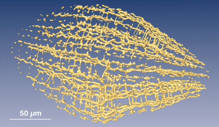
Figure 129 shows a virtual slice and the corresponding schematic drawings. Individual cells and organelles inside the cells are clearly visible in the enlargement of a section (Figure 129d). In the reproduction scheme used, darker corresponds to higher density. The white dots showing up at the junction of cells correspond to essentially zero density, i.e. to gas. More detailed 3D scrutiny, as in Figure 130 [2] shows that the voids form a network. This network of air space is the major finding of this investigation. It may be an important actor in plant life, by first storing and then, at the onset of the germination process, quickly distributing oxygen. In addition, it could serve for rapid water transport to the various seed tissues and organs during imbibition. A noteworthy aspect is that this air space network could not be reliably identified using the standard approach of fixation, dehydration, embedding, microtome slicing, colouring, and optical microscope observation. The reason is that slicing requires the various liquids, in particular the polymer embedding agent, to permeate the sample. Due to the imperviousness of the seed coat, this is impossible unless incisions are deliberately made. In contrast, the X-ray tomography approach is completely non-destructive. Another question worth addressing is whether high energy (typically 21 keV) X-rays are the right probe for investigating a minute (0.4 mm), low-absorption object such as this seed. The answer is that they provide a unique combination of the desired resolution (0.3 µm pixel size was used here), field of view (2000 x 2000 pixels) and depth investigation capacity (the whole sample). While the irradiation dose involved in this phase tomography investigation is high, it is less high than in attenuation contrast tomography. The investigation was made possible by the fact that no visible change in the morphology of the seed occurred, whereas irradiation usually leads to distortion in wet samples.
 |
|
Fig. 130: Network of voids in the seed. |
In conclusion, holotomography has made it possible to obtain the first 3D images and renditions of an autonomous living object, a small seed, at the sub-micrometre scale and in a non-destructive manner. The air-space network evidenced for the first time in this study may play a central role in the germination process.
References
[1] P. Cloetens, W. Ludwig, J. Baruchel, D. Van Dyck, J. Van Landuyt, J. P. Guigay, and M. Schlenker, Appl. Phys. Lett. 75, 2912 (1999).
[2] The image can be rotated in the supporting material to the original publication, http://www.pnas.org/cgi/ content/full/0603490103/DC1.
Principal Publication and Authors
P. Cloetens (a), R. Mache (b), M. Schlenker (c), and S. Lerbs-Mache (b), Proc. National Acad. Sci., (USA),103, 14626-14630 (2006).
(a) ESRF
(b) Laboratoire Plastes et Différentiation Cellulaire, UJF/CNRS, Grenoble (France)
(c) Département Nanosciences, Institut Néel, CNRS/UJF/INPG, Grenoble (France)


