- Home
- Users & Science
- Scientific Documentation
- ESRF Highlights
- ESRF Highlights 2006
- X-ray Imaging and Optics
X-ray Imaging and Optics
Introduction
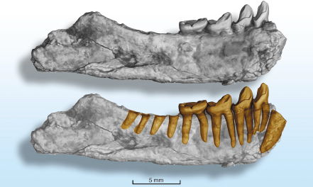
X-ray imaging and microanalysis techniques cover a wide variety of scientific topics ranging from materials science and engineering to life sciences and cultural heritage. The evolution of these techniques is science-driven: a good example is the success achieved when establishing a link with paleontological research, and adapting the techniques to the needs of this community [1,2]. Their results are exemplified by the three-dimensional images of a fossil shown in Figure 123.
 |
|
Fig. 123: Mandible of a small fossil strepsirrhine primate from the Late Eocene of Thailand, Muangthanhinius siami. Non-destructive three-dimensional observation of the teeth roots (ID19, pixel size 10 µm) gave clues that let us better understand the morphology of the teeth crown. The image analysis allowed a modification of the attribution of this fossil, originally considered as a putative ancestor of lemurs, and now reattributed to an extinct group of primates, the Adapiforms [3]. Image courtesy P. Tafforeau. |
The present chapter clearly shows the variety of scientific subjects investigated in connection with imaging and microanalysis techniques: it goes from preclinical radiotherapy studies to optics for nanofocussing, and includes applications to biomedical and biological science, materials science, environmental and cultural heritage topics.
The selected biomedical research articles comprise both radiotherapy and X-ray imaging. The present number of treatments for cancerous brain tumours, and their efficiency, is unfortunately quite poor. It is therefore particularly important to develop synchrotron radiation based research aimed at treating brain tumours. Two contributions, one on survival curves using synchrotron stereotactic radiotherapy (SSR), and another on the sparing of healthy tissues when using microbeam radiation therapy (MRT), go in this direction. A report on the time response to inhaled histamine, studied by synchrotron radiation computed tomography (CT) using Xe as a contrast agent and the K-edge subtraction method, provide information on the complex changes in conductance of the respiratory system, important for the understanding and treatment of asthma.
Intense research is being performed using imaging techniques on biological materials. Holotomography with very high spatial resolution allowed the investigation of an Arabidopsis seed, and showed the existence of a three-dimensional air space network, which may play a key role in the germination process. In the second article, spatially-resolved diffraction was used to map the texture of human dental enamel.
Application of these techniques to materials science continues to provide many original results. Microstructure formed during the growth process controls a substantial fraction of a material’s properties. The observation of the solid-liquid interface during directional solidification is essential for studying the dynamical phenomena involved in microstructure formation under controlled conditions. A unique experimental setup, allowing the combination of synchrotron X-ray radiography (SXR) and synchrotron white beam X-ray topography (SWBXT), has been developed. These two imaging techniques provide complementary information on the growth of Al-based alloys: both the evolution of the microstructure morphology and the orientation and strain field within the growing solid are accessible in this way. The final materials-related contribution is a µm-scale spatially-resolved investigation of Co impurities in ZnO which allows an identification of the mechanism inducing ferromagnetism in this material.
Imaging and microspectroscopy are being increasingly used to investigate environment and cultural heritage subjects. The highlighted contributions start with a note explaining the mechanism of blackening of the red paintings of Pompeï. The evolution from Fe3+ to Fe2+ under conditions close to those in magmas during their ascent from the Earth’s mantle to the surface, before volcanic eruption, furthers our knowledge of the whole process. Finally the importance of the use of phase contrast to identify insects or plants within opaque fossil amber is pointed out, and three-dimensional images of the fossils identified in this way are shown.
This chapter also includes a note on X-ray optics metrology, which shows the present achievements and emphasises the importance of refined metrology to meet future requirements of nanofocussing optics.
X-ray imaging plays a key role within the planned ESRF Upgrade Programme. The present work on X-ray imaging shows the importance of responding to the increasing demand from new scientific communities and gains from the continuous improvements to the techniques. A further effort to improve the spatial resolution to reach the few tens of nm range, for both biological (such as micro-vascularisation of the brain, trace elements in cells) and materials science (such as initiation of cracks) topics, is crucial. It implies building a new, devoted and optimised, long nanoimaging beamline to complement the set of techniques already established within the X-ray Imaging group.
J. Baruchel
References
[1] J-Y. Chen et al., Science 312, 1644-1646 (2006).
[2] R. Macchiarelli et al., Nature 444, 748 - 751 (2006).
[3] L. Marivaux et al., Am. J. Phys. Anthropol. 130, 425 (2006).


