X-ray imaging
Synchrotron-based X-ray imaging techniques use a broad range of physical and chemical contrast mechanisms. Their ‘resolution’ can be seen in three dimensions: spatial, temporal and compositional/structural. Synchrotron radiation-based high resolution X-ray imaging is an invaluable tool for the study of complex systems, leading to substantial scientific progress. The highlights published in the present chapter show the wide variety of topics studied at the ESRF. Imaging at the ESRF is now approaching spatial resolutions in the few tens of nm range, well below the cell dimensions, and chemical sensitivities allowing the detection within the cell of a single nanoparticle consisting of only a few thousand atoms (ID22). In the first section on biology and biomedical applications, this is highlighted by the contribution on new opportunities for bioimaging and biodelivery, which have potential applications in diagnostics and therapy of diseases such as cancer. Two phase-contrast imaging developments are also presented; both aim to improve medical diagnosis. Grating interferometry tomography allows discrimination between soft tissues such as white and grey matter in a human cerebellum sample (ID19), and phase contrast analyser-based tomography permits differentiation between osteoarthritic and healthy human cartilage samples (ID17). The last paper of this section deals with the tolerance of arteries to microplanar X-ray beams, a study that is required before the new “micro-radiation therapy” (MRT) protocol can be applied to the treatment of cancerous tumours.
High resolution is also important for materials science applications, where “local” diffraction maps are collected at the micrometre level. Two examples are presented here. The growth process of quantum-well based devices for optoelectronics can be improved using the information gained from their imaging (ID22), and superconductivity is associated with the connectivity over large distances within the nanostructure for oxide-based superconductors (ID13).
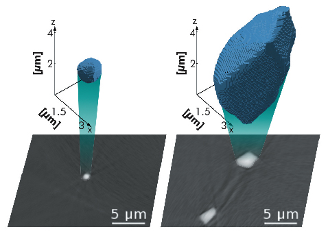
Works for the upgrade beamline UPBL4 NINA (Nano Imaging and Nano-Analysis) officially started at the end of 2010. This two-branch beamline will provide complementary techniques at the nano-scale for the study of both biological and materials science samples. Nano-imaging covers new application areas and has been pioneered by the pilot project ID22NI. This is exemplified by the study of the growth of grain boundary voids during the high-temperature creep of metals. The usual model considers this process similar to the growth of a hole in a nonlinearly viscous solid. Fast high-energy tomography shows that the real growth rates in copper are higher than the prediction of this model by a factor of about 40. Nano-tomography reveals that voids have a spherical shape in the initial undeformed state and they become facetted during creep (Figure 123). This observation and the large scatter of growth rates suggest that dislocation glide significantly contributes to void growth [1].
 |
|
Fig. 123: Submicrometre resolution reconstructions of void shape. a) A spherical shape is characteristic of the initial, recrystallised state. b) Larger voids become faceted after 4% of creep deformation at 723 K (Image copyright 2010, reprinted from [1] with permission from Elsevier). |
X-ray imaging is increasingly being used for Cultural Heritage investigations. A combination of fluorescence (µXRF) and absorption (µ-XANES) on ID21 allowed characterisation of the opacification processes in Egyptian and Roman glass. This study highlighted the remarkable know-how of the craftsmen of those periods. Synchrotron radiation based imaging makes it possible to reach elusive information that can completely change our understanding of a palaeontological subject. One study shows that differences existed between the Neanderthal dental development and our own, and that a long childhood is specific to the evolution of our own species (ID19). This investigation is an example of the kind of research carried out in the “Palaeontology Facility”, which aims to provide the palaeontological community with specially designed facilities. It will involve the refurbishing of long imaging beamlines and a specialised computing centre. The Palaeontology Facility will involve collaboration among many internationally renowned groups, and it will reinforce the leading role of the ESRF for this scientific activity. Already this effort is producing important new results, such as the very recent evidence about the diet of one of the most important group of ammonites, distant relatives of squids, octopuses and cuttlefish, which sheds a new light on why they became extinct 65.5 million years ago [2].
New science often relies on technical developments: the ESRF staff devote a substantial part of their research to achieving these improvements, as exemplified by two contributions to the present highlights. The first account describes a newly developed wavelength-dispersive spectrometer (ID21) with performance that significantly surpasses that of a solid state detector. In the second article, real-time characterisation is achieved for the evolution of the roughness of silicon surfaces upon ion erosion, this being important for the production of X-ray optical elements. High-resolution synchrotron X-ray imaging (and in particular microtomography) is increasingly being required for industrial, proprietary studies. This very positive feature raises the issue of the beamtime/personnel for these highly oversubscribed beamlines, and the need to integrate this aspect in the X-ray imaging development strategy at the ESRF.
J. Baruchel
References
[1] K. Dzieciol, A. Borbély, F. Sket, A. Isaac, M. Di Michiel, P. Cloetens, Th. Buslaps and A.R. Pyzalla, Void growth in copper during high-temperature power-law creep, Acta Materiala 59, 671-677 (2011).
[2] I. Kruta, N. Landman, I. Rouget, F. Cecca and P. Tafforeau, The Role of Ammonites in the Mesozoic Marine Food Web Revealed by Jaw Preservation, Science 331, 70-72 (2011).
partners


European Synchrotron Radiation Facility - 71, avenue des Martyrs, CS 40220, 38043 Grenoble Cedex 9, France.