- Home
- Users & Science
- Find a beamline
- Collaborating research group beamlines
- BM32 - IF - InterFace Beamline, French CRG
- BM 32 Featured Articles
- In situ GISAXS study of the self-organized growth of Co on Pt/W(111) faceted surface
In situ GISAXS study of the self-organized growth of Co on Pt/W(111) faceted surface
In situ GISAXS study of the self-organized growth of Co on Pt/W(111) faceted surface
F. Leroy, G. Renaud, T.E. Madey, A. Letoublon, R. Lazzari, T. Schülli
This page is under construction...
T.E Madey team has recently shown that a single monolayer of Pt adsorbed on an atomically rough W(111) surface undergoes a huge morphological change upon annealing above 700K : a complete faceting of the surface in the form of ordered triangular nanopyramids exposing {211} facets is thermodynamically driven via surface energy anisotropy. The growth of nano-pyramids is kinetically limited. Near field microscopy have been extensively used on this system as well as low energy electron microscopy, but both techniques are limited respectively by the temperature range and the limited resolution for a characterization of the very beginning of nanofaceting. The challenge gets even more difficult when the study focuses on the self-organized growth of cobalt on such a highly patterned substrate.
 |
 |
|
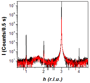
Fig. 1 In-plane scans unambiguously show that all the Pt deposited is pseudomorphic, as shown in this example: a h-scan along h00 before and after deposition of 1 ML of Pt. |
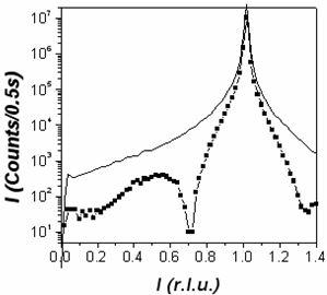
Fig. 2 : (20L) L-scan before and after 1 monolayer of Pt deposit on the W(111) surface. A strong interference effect is clearly visible. Quantitative analysis of 5 integrated CTRs should yield the exact Pt epitaxial site and distance. |
We have combined in situ Grazing Incidence Small Angle X-Ray Sattering (GISAXS), Grazing Incidence X-Ray Diffraction (GIXD), X-ray reflectivity and ex situ STM and AFM to probe, during growth, the faceting of the surface as well as the growth of Co nanostructures. Much effort has been made to nucleate few nanometers wide ordered nano-pyramids with the sake of using them as a template for self-organized growth of cobalt.
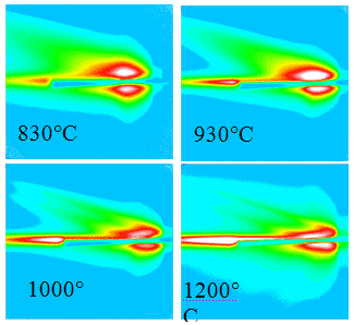 |
Fig. 3 : GISAXS image of the faceted surface at different temperatures. The sample is on the left, the surface normal being horizontal. |
W(111) substrates have been prepared by repetitive annealing in oxygen (1x10⁻⁷ Torr) at 1700K, followed by flashes up to 2600K in order to get ride of carbon. The absence of surface impurity was checked by AES and X-ray diffraction (tungsten carbides and oxides have very clear signatures by GIXD). The experimental set-up contains 4 Omicron EFM4 e-beam bombardement deposition sources, 2 for Pt and 2 for Co, calibrated first by a Quartz Crystal Microbalance, next by anti-Bragg X-ray intensity oscillations and AES. First, we have characterized the structure of the Pt/W(111) interface by quantitative CTR measurements on the planar surface to deduce the epitaxial relationship between perfectly pseudomorphic Pt, see Fig. 1, and W [Fig. 2]. Then short annealings of a few minutes at growing temperature by step of 50K have been performed to probe the faceting process. Characterization by GISAXS and GIXS of the scattering rods of facets have revealed the internal structure of pyramids and their morphology. Unfortunately quantitative results have only been obtained on quite large pyramids (20-100nm) and the smallest pyramids as well as cobalt deposit were only qualitatively studied.
GISAXS and GIXS on large facets
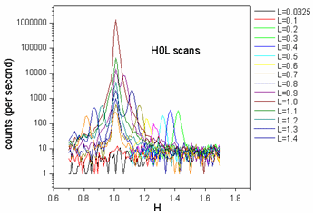
GISAXS measurements confirmed the threefold symetry of the pyramids and the large predominance of {211} facets as compared to {110} facets. Upon annealing, pyramids grow in size as shown by the scattering rods that narrow, intensify and get closer to the origin of the reciprocal space [Fig. 3]. The doubling of rods as the incident angle get closed to the critical angle reveals the expected effects of the DWBA which takes into account a ‘beam multiplier’ effect of the surface near the critical angle. Quantitative measurements of the CTR of {211} facets should yield the epitaxial relationship between Pt and W(211), information on possible intermixing between W and Pt on these faces as well as on the strain and the of these large pyramids [Fig. 4].
 |
 |
|
Fig. 4 h-scans along for different L values revealing rods of scattering by {211} facets |
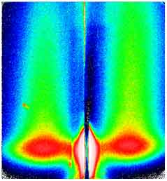
Fig. 5: GISAXS image of small correlated Nano-pyramids separated by 12 nm. The sample is at bottom and the vertical direction is perpendicular to the surface. The specular beam is hidden by a beam-stop |
GISAXS and GIXS on small pyramids and Co deposit
The temperature range for the nucleation of a few nanometer wide (5-10nm) ordered pyramids is very narrow and, although this temperatures are now very well known, a full quantitative characterization of the nano-facetted surface have not been obtained, but only qualitative results. GISAXS measurements have revealed correlated 5-10 nm-wide pyramids, exposing {211} facets [Fig. 5]. On the cobalt deposit upon small pyramids, GISAXS pattern seems to remain unchanged upon 3ML of Cobalt.


