- Home
- Users & Science
- Scientific Documentation
- ESRF Highlights
- ESRF Highlights 2000
- Life Sciences
- Structure Determination using the Anomalous Signal from Sulphur Atoms
Structure Determination using the Anomalous Signal from Sulphur Atoms
Crustacyanin is a chromophore binding protein found in lobster carapace. Crystals of the C1 subunit of the protein are of excellent quality and diffract to atomic resolution. However, despite many efforts using both molecular replacement and multiple isomorphous replacement techniques, the structure determination appeared to be insoluble. Furthermore, in the absence of suitable constructs, it was not possible to produce a Selenomethionine substituted protein for MAD experiments. An unusual approach was therefore undertaken based on the knowledge that the protein possesses three di-sulphide bonds. The positions of these sulphur atoms, intrinsic to the dimeric native protein, were located using the anomalous signal from sulphur. These sulphur positions were then used in a new ab initio phasing procedure to produce phases to atomic resolution and a readily interpretable electron density map.
Data were collected from a single crystal on BM14, with a wavelength of 1.77 Å. At this wavelength the expected f" signal from the sulphur is 0.75e-. The expected signal for 12 sulphurs in 362 residues in the asymmetric unit is of the order of 1%. 560 degrees of data were collected, which gave 100% completeness to 2.5 Å, with a multiplicity of 21.3 and an overall Rsym = 4.6%.
SnB [1], a direct methods program, was used to determine the sulphur atomic positions from the anomalous signal. Six positions were found, which corresponded to one of the two sulphurs present in each disulphide bridge. These six sites exhibited the non-crystallographic symmetry expected for the dimeric arrangement of molecules in the crystallographic asymmetric unit. Refinement of these sulphur positions against the data gave encouraging statistics. Maximum likelihood residual maps revealed six more positions corresponding to the second sulphur atoms in each disulphide bridge. All 12 sulphur sites were then refined to give occupancies of 1.0 and a Figure of Merit of 0.10 for centric reflections and 0.36 for acentric reflections to a resolution of 2.5 Å. However, no further progress was achievable at this stage using conventional phasing programs.
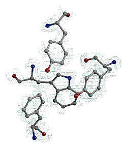
The twelve sulphur sites were subsequently used with a high-resolution data set (1.15 Å), collected from the same crystal with a completeness of 100% and an overall Rsym = 6.0%. Using the ab initio phasing program ACORN [2], the twelve sulphurs were used to calculate an initial phase set and these phases were refined and extended to produce a map at 1.15Å which was readily interpretable. Figure 8 shows part of the electron density map section with various aromatic residues clearly visible.
 |
Fig. 8: Part of the electron density map of crustacyanin.
|
Although using the anomalous signal from sulphurs has already been demonstrated to be a successful phasing tool for small proteins [3], this is the first time that it has been demonstrated to work for an unknown protein structure. Future work will explore the general utility of this method for phasing macromolecular structures.
References
[1] R. Miller, S.M. Gallo, H.G. Khalak and C.M. Weeks, J. Appl. Cryst. 27, 613-621 (1994).
[2] J. Foadi, M.M. Woolfson, E.J. Dodson, K.S. Wilson, Y. Jia-xing, and Z. Chao-de, Acta Cryst. D56, 1137-1147 (2000).
[3] Z. Dauter, M. Dauter, E. de la Fortelle, G. Bricogne and G.M. Sheldrick, J. Mol. Biol. 289, 83-92 (1999).
Authors
E. Gordon (a), S. McSweeney (a), G. Leonard (a) and P. Zagalsky (b).
(a) ESRF
(b) Royal Holloway College (UK)


