- Home
- Users & Science
- Scientific Documentation
- ESRF Highlights
- ESRF Highlights 2002
- Soft Condensed Matter
- Supramolecular Organisation of Biomimetic Nanotubes
Supramolecular Organisation of Biomimetic Nanotubes
Controlled self-assembly of molecules into well-defined nanoscale structures is a challenging task, which has wide-ranging applications in biotechnology and materials science. These nanoscale structures can be realised by using naturally occurring proteins such as tobacco mosaic virus (TMV) [1]. Biological entities like viruses or microtubules, are perfectly monodisperse thanks to the perfection achieved by mother nature in protein self-assembly. In general, such level of perfection is hard to obtain in artificial systems. However, an alternative route has become feasible, based on synthetic molecules that can be programmed to self-organise in a tailored fashion. The design of such systems requires the understanding of the interrelationship between the molecular structure and the self-assembly process to nanostructures [2]. New strategies have developed using inorganic materials, lipids and amphiphilic compounds. One of the most promising approaches is based on the de novo synthesis of oligopeptide that self-organises in well-defined fibres.
 |
|
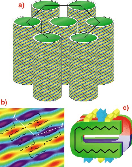
Fig. 21: a) hexagonal packing of the nanotubes, b) close-up of electron density and c) molecular conformation within the fibre. |
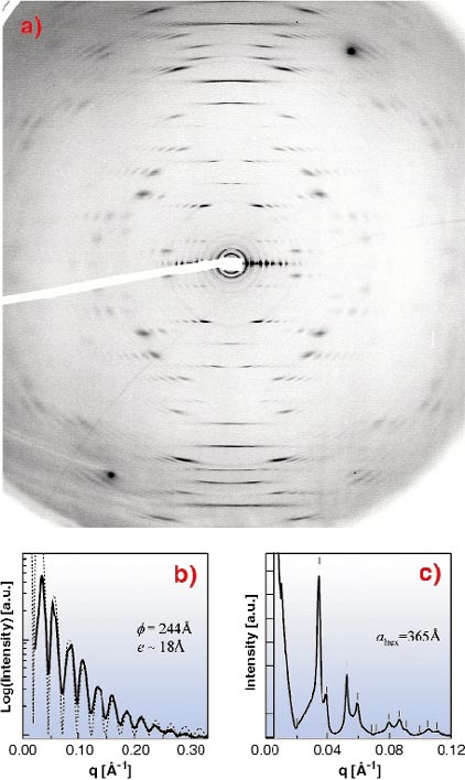
In this context, the Lanreotide octapeptide developed by Beaufour-Ipsen laboratory that forms a therapeutic gel (autogel®) is an illustrative example. At a lower Lanreotide concentration than in the gel, monodisperse nanotubes are spontaneously formed (Figure 21). This work concerns the elucidation of supramolecular and molecular organisation by small and wide angle X-ray scattering (SAXS and WAXS, respectively), carried out on ID02, and the complementary techniques of electron microscopy and vibrational spectroscopies. The exceptionally well-aligned WAXS pattern (Figure 22) reveals that the organisation within the nanotubes wall is crystalline with low mosaicity (< 0.1°). This diffraction pattern acquired with a Lanreotide derivative can be unambiguously interpreted in terms of a 2D curved crystal and it is quite similar to the pattern recorded for Tobacco Mosaic Viruses [1]. The Patterson function of the nanotubes wall, calculated from the diffuse scattering, indicates a 20.7 Å alternation of aliphatic and aromatic residues that form beta-amyloid like fibres. The nanotubes are constituted by 26 of these fibres and consequently their diameter is very monodisperse as evidenced by SAXS (Figure 22). High-resolution SAXS further reveals the hexagonal packing of these tubes.
 |
|
Fig. 22: (a) Fibre diffraction of the nanotube, (b) SAXS demonstrating the tube monodispersity and (c) high-resolution SAXS showing the hexagonal packing of the nanotubes. |
A multidisciplinary approach involving high-resolution X-ray fibre diffraction and molecular spectroscopies (FTIR, FT-Raman) is essential to solve the Lanreotide nanotube structure at different hierarchical levels. The complete picture demonstrates a systematic segregation between aliphatic and aromatic residues at the origin of the nanotube organisation. High-resolution small-angle X-ray scattering revealed the nanotube sizes (diameter, wall thickness) and their packing. The controlled supramolecular organisation will be a promising route in the future for designing de novo molecules that can self-assemble into nanotubes with tuneable diameter.
References
[1] A. Klug, Angew. Chem. Int. Ed. Engl. 22, 565-636 (1983).
[2] J.-M. Lehn, Angew. Chem. Int. Ed. Engl. 29, 1304-1319 (1990).
Authors
F. Artzner (a), C. Valery (a), M. Paternostre (a,b) T. Narayanan (c)
(a) UMR 8612 CNRS, Faculté de pharmacie de Chatenay-Malabry (France)
(b) URA 2096 CNRS, CEA Saclay (France)
(c) ESRF


