- Home
- Users & Science
- Scientific Documentation
- ESRF Highlights
- ESRF Highlights 2003
- Industrial and Applied Research
- In situ Characterisation of Aluminium Foams using X-ray Microtomography
In situ Characterisation of Aluminium Foams using X-ray Microtomography
Metal foams can now be produced with Ni or Al alloy in open or closed-cell morphology. Open cell morphologies are achieved by coating a polymer foam and also by replicating salt preforms. These two techniques allow a very good control of the structure of the foam. Closed-cell morphology is mainly obtained with aluminium alloys and whatever the process (liquid route or powder route) the control of the foaming process and thus of the final structure is not easy. This may lead to foams which present an inhomogeneous structure. Classical mechanical models do not take into account such a structural heterogeneity, thus the question of the influence of inhomogeneity on the mechanical properties is still an open question. Owing to the good contrast between air and the constitutive materials, X-ray absorption techniques can be used to image the structure. For example, X-ray radiography has been used to study the foaming of metal foams, X-ray tomography was successfully used to characterise bone, polymer foams and also metal foams at various resolutions.
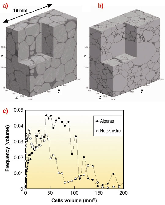
The aim of the experiments was to characterise the 3D structure of the various aluminium foams that are available and to study their in situ behaviour during compression and tensile tests using a specific device [1]. They were performed at beamine ID19 at energy ranging from 18 to 25 KeV and with 10-30 micrometre optics. 900 projections were recorded on the 1024x1024 FRELON camera. From the data obtained, 3D quantitative analysis of the various aluminium foams allowed a comparison of four kinds of closed cell aluminium foams, produced by various processing routes (IFAM, Alporas, NosrkHydro, Formgrip), in terms of cell size distribution (even when they are not well closed), aluminium phase distribution and connectivity of the cells. Figure 153 presents two closed cell aluminium foams and their cell size distribution. This clearly shows the quite good homogeneity of the Alporas foam, whereas the NosrkHydro foam presents some large cells (volume ~ 150 mm3) that are visible on the 3D rendering.
 |
|
Fig. 153: a) 3D rendering of Alporas foam; b) 3D rendering of NosrkHydro foam; c) Cell size distribution obtained from 3D granulometry. |
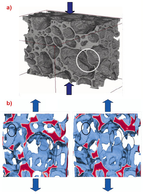
In situ experiments were also performed in order to clearly identify the mechanical mechanisms that occur during the compression (closed cell foam) and tensile testing (open cell foam). The main mechanisms are: plastic buckling of cell walls (see Figure 154a) when the constitutive material is quite ductile (IFAM, Alporas) which may be followed by rupture at higher strain for less ductile foams (FormGrip). NorskHydro foam presents rupture of the cell walls due to the brittle nature of the constitutive material. Concerning the open-cell foams, the tensile behaviour of Ni [2] and pure aluminium foam investigated allows us to draw conclusions concerning the damage mechanisms: Figure 154b shows clearly that a pure aluminium foam undergoes plastic straining of struts and also rupture of the struts. Furthermore the strain does not seem to be homogeneous along the sample.
 |
|
Fig. 154: a) Damage mechanism in a closed cell Al foam (NorskHydro) during in situ compression test. Plastic buckling of a cell wall is indicated with the circle (width of the specimen ~ 15 mm); b) In situ damage mechanism in a pure Al foam (salt replication technique made at EPFL) during in situ tensile testing. Two successive stages are represented. Continuous black circle shows plastic straining of a strut and discontinuous black circle rupture of struts (width of the specimen ~ 1 mm). |
This work is still in progress and some questions remain without definitive answers. Indeed, if the main mechanism observed at the cellular scale are now well understood, the correlation of what is locally observed and the macroscopic behaviour of the foam is not well established. 3D density mapping combined with 3D strain mapping and FEM analysis from the tomography data will certainly be useful to answer this question.
References
[1] J-Y. Buffière, E. Maire, P. Cloetens, G. Lormand, R. Fougères., Acta Met , 47, 1613 (1999).
[2] T. Dillard, F. Nguyen, S. Forest, Y. Bienvenu, J.D. Bartout, L. Salvo, R. Dendievel, E. Maire, P. Cloetens, C. Lantuéjoul, in "Cellular Metals: manufacture, properties, applications", J. Banhart, N. Fleck, A. Mortensen Eds, Verlag MIT Publishing, 301 (2003).
Principal Publications and Authors
A. Elmoutaouakkil (a), L. Salvo (a), E. Maire (b), G. Peix (b), Adv. Eng. Mat., 4(10), 803 (2002); E. Maire (b), A. Elmoutaouakkil (a), A. Fazekas (a), L. Salvo (a), MRS bull., 28(4), 284 (2003).
(a) GPM2, INP Grenoble (France)
(b) GEMPPM, INSA Lyon (France)


