- Home
- Users & Science
- Scientific Documentation
- ESRF Highlights
- ESRF Highlights 2003
- Industrial and Applied Research
- Evaluation of 2D Radiographic Texture Analysis for Assessing 3D Bone Micro-architecture
Evaluation of 2D Radiographic Texture Analysis for Assessing 3D Bone Micro-architecture
The prevalence of osteoporosis increases significantly with life expectancy in industrial countries. Osteoporosis is a bone disease characterised by low bone mass and structural deterioration of bone tissue, leading to bone fragility and an increased susceptibility to fractures of the hip, spine, and wrist. The clinical evaluation of osteoporosis relies on Dual X-ray Absorptiometry (DXA) which provides an estimation of the Bone Mineral Density (BMD) typically on the spine and on the hip. Unfortunately, an important overlap of BDM values is observed in patients with and without fractures, and BMD alone is not sufficient to predict individual fracture risk. Another factor to take into account in the biomechanical resistance of bone, is its micro-architecture. The latter has conventionally been investigated using histomorphometry, but during the last decade tomographic techniques have been more and more employed [1]. Though, due to the requirements in terms of spatial resolution this investigation is limited to in vitro examinations on bone biopsies.
Standard X-ray radiography associated with texture analysis has been proposed by different teams to get in vivo architectural information. Although the information delivered by a single radiograph is somehow limited, the discrimination of osteoporotic patients and healthy subjects has been demonstrated in clinical studies [2]. However, the most appropriate texture parameter(s) and the optimal imaging conditions for the description of bone micro-architecture are still unknown.
The purpose of this collaborative project (CEA-LETI, CREATIS, ESRF, CHU Nîmes and Lille) was to develop a method for evaluating the relationships between 3D micro-architecture parameters and 2D texture parameters, and for optimising the conditions of radiographic image acquisition. It is expected that such conditions could be implemented on new generations of bone densitometers such as the LEXXOS, developed by CEA-LETI and DMS, based on a 2D digital radiography flat panel technology.
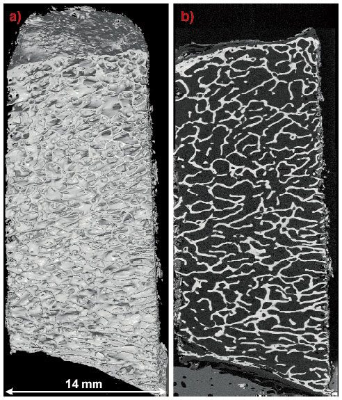
A procedure based on the use of reference images of representative human bone samples acquired using 3D-synchrotron X-ray microtomography was developed at the ESRF. Thirty-three cylindrical samples (diameter 14 mm) were taken in calcaneus and femoral neck, from cortical to cortical to be close to in vivo conditions. Imaging was performed on the microtomography setup developed on beamline ID19. To have a sufficient field of view, a spatial resolution of 15 µm was used, and scans at different heights were acquired to encompass the entire sample (total height: 30 to 40 mm). Figure 158 shows a 3D display of a reconstituted entire sample as well as one 2D slice.
 |
|
Fig. 158: Calcaneus bone sample (mo10) imaged using synchrotron radiation microtomography on beamline ID19 at the ESRF (voxel size: 15 µm): (a) 3D display of the reconstituted entire sample (b) 2D vertical slice. |
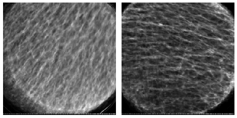
These digital three-dimensional images were further used both for calculating three-dimensional quantitative architecture parameters of trabecular micro-architecture, and simulating realistic X-ray radiographs under different acquisition conditions using the Sindbad software (CEA-LETI) [3]. Figure 159 illustrates simulated radiographs of two samples at a spatial resolution of 50 µm.
 |
|
Fig. 159: Radiographs simulated from the 3D discrete images of two entire calcaneus samples (pixel size: 50 µm). left: same sample as Figure 158, right: sample moca4. |
Texture analysis was then applied to these simulated 2D radiographs using software developed at CREATIS including a large variety of methods (co-occurrence, spectrum, fractal) and delivering more than one hundred parameters per image. The set of texture parameters was sequentially reduced according to different criteria to find the most relevant ones to three dimensional architecture. Finally, six texture parameters significantly correlated to bone micro-architecture parameters were selected. The relevance of these parameters will be confirmed by further tests on physical radiographs of the samples acquired in different experimental conditions (energy range and spatial resolution). The implementation of such a technique on a bone densitometer would allow to supplement the standard BMD with an architectural index for expected better discrimination with respect to the fracture risk.
References
[1] F. Peyrin, M. Salome, S. Nuzzo, P. Cloetens, A.M. Laval-Jeantet, J. Baruchel, Cellular and Molecular Biology, 46(6): 1089-1102, (2000).
[2] L. Pothuaud, E. Lespessailles, R. Harba, R. Jennane, V. Royat, E. Eynard, C.L. Benhamou , Osteoporosis Int., 8, 618-625, (1998).
[3] R. Guillemaud, J. Tabary, P. Hugonnard, F. Mathy, A. Koenig, A. Glière, PSIP, (2003).
Principal Publication and Authors
F. Peyrin (a,e), L. Apostol (a, e), E. Boller (e), O. Basset (a), C. Odet(a), S. Yot (a), J. Tabary (b), J.M. Dinten (b), V. Boudousq (c), P.O. Kotzki (c), X. Marchandise (d), SPIE Med. Imaging, 2004.
(a) CREATIS (France)
(b) CEA-LETI (France)
(c) CHU Nimes (France)
(d) CHU Lille (France)
(e) ESRF


