- Home
- Users & Science
- Scientific Documentation
- ESRF Highlights
- ESRF Highlights 2003
- Soft Condensed Matter
- A Novel Method to Investigate Gradient Multilayers on Multiple Length Scales
A Novel Method to Investigate Gradient Multilayers on Multiple Length Scales
Nanometre-sized noble metal clusters are of great technological and scientific importance being widely used in biomedical analysis, e.g. DNA sequencing [1], due their special optical properties being distinctly different from bulk material. This results from plasmon resonances occurring as a consequence of the confinement of the free electron gas inside the nanometric noble metal cluster. Due to the electromagnetic dipole interaction even a single molecule in the close vicinity of a cluster can be detected and characterised using optical spectroscopy.
The optical properties of the cluster layer strongly depend on their three-dimensional shape and in-plane arrangement. Typical systems consist of a layer of nanometre-sized noble metal clusters on top of a polymer film. Especially for property optimisation, it is necessary to vary the structure and morphology of the cluster layer and to produce laterally-heterogeneous multilayer systems. Hence, we investigated the morphology and structure of a lateral gradient layer of nanometre-sized gold clusters on top of a polystyrene (PS) layer on a Si mirror substrate.
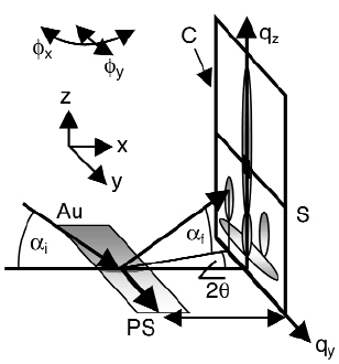
Our novel approach is based on a combination of grazing-incidence small-angle X-ray scattering (GISAXS) with a microbeam available at beamline ID13 (giving rise to the µGISAXS method [2]). Here, a 5 µm monochromatic X-ray beam impinges under a low angle onto the sample surface (Figure 81). In this way, we were able to spatially resolve the morphology of the nanometre-sized gold cluster layer along the gradient on a micrometre scale.
 |
|
Fig. 81: Geometry of the microbeam grazing-incidence small-angle X-ray scattering (µGISAXS) experiment at ID13. The monochromatic beam ( |
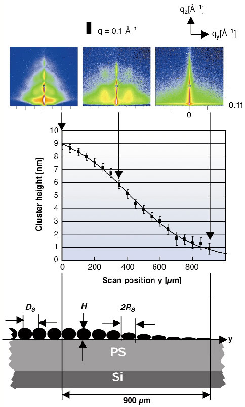
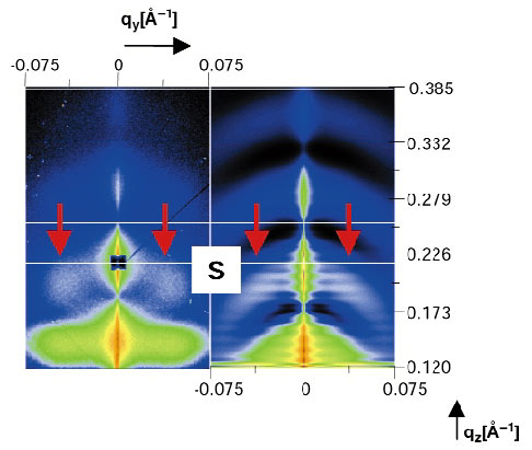
In Figure 82 we present typical mGISAXS patterns. The gradient was scanned with a step size of 50 µm. The µGISAXS patterns clearly change along the gradient. Analysing the full two-dimensional pattern yields the arrangement and the three-dimensional shape of the gold clusters. To do so we simulated the µGISAXS pattern (program IsGISAXS by R. Lazzari) for each scan point y using a detailed model assumption for the gold cluster layer and including the Si-substrate and the PS-layer. Figure 83 shows a typical example. From the position of the side maxima one can deduce the in-plane radius and distance of the gold clusters. It turned out that these two parameters stay constant along the gradient (RS = (10.6 ± 5.3) nm, DS = (14 ± 10) nm). Assuming an ellipsoidal model for the cluster shape, their height can be deduced from the position of the minima at each scan point y. The middle inset in Figure 82 shows the cluster height as a function of y. It follows a hyperbolic tangent profile.
 |
|
Fig. 82: Upper part: µGISAXS patterns recorded at y = 0, 350, 900 mm respectively. Middle part: Change in gold cluster height as a function of the scan position y. The solid line shows a fit of the experimentally-determined cluster height using a hyperbolic tangent function. Lower part: Schematic side view of the gold cluster arrangement along the gradient. DS denotes their in-plane distance, 2RS their in-plane diameter. The polystyrene (PS) film has a nominal thickness of 40 nm. The substrate is Si. The gradient extends over 900 µm. |
 |
|
Fig. 83: Comparison of data (left) and simulation (right). The data show two principal features: minima along qz and side maxima at finite qy and qz. The minima (white lines) indicate the cluster height, while from the side maxima (red arrows) the in-plane radius RS and the in-plane distance DS can be derived. The best fit is obtained using an ellipsoidal model for the gold clusters, where only their aspect ratio changes as a function of y. |
To conclude, µGISAXS was shown to be a powerful non-destructive method to scan and thus to reconstruct the structure and morphology of laterally inhomogeneous multilayer systems on a micrometre scale. The gradient of ellipsoidal nanometre-sized gold clusters on PS/Si is characterised by one single parameter: the cluster height. Hence their aspect ratio changes along the gradient. We plan to extend the beam size to the sub-micrometre range in the near future.
References
[1] B. Dubertret, M. Calame and A. J. Libchaber, Nature Biotech. 19, 365 (2001).
[2] P. Müller-Buschbaum, S.V. Roth, M. Burghammer, A. Diethert, P. Panagiotou, C. Riekel, Europhys.Lett. 61, 639 (2003).
Principal Publication and Authors
S.V. Roth (a), M. Burghammer (a), C. Riekel (a), P. Müller-Buschbaum (b), A. Diethert (b), P. Panagiotou (b), H. Walter (c), Appl. Phys. Lett. 82, 1935 (2003).
(a) ESRF
(b) Technische Universität München, Garching (Germany)
(c) November AG, Erlangen (Germany)


