- Home
- Users & Science
- Scientific Documentation
- ESRF Highlights
- ESRF Highlights 2003
- Surface and Interface Science
- Super-cell Crystallography of Self-organised Deposits
Super-cell Crystallography of Self-organised Deposits
Self-assembly consists of the spontaneous formation of nanostructures in the first stages of growth on a surface, by tailoring instabilities such as the Stranski-Krastanov growth mode. Self-assembly is currently of high interest because of its ability to produce nanostructures in a single-run process that are smaller (5-50 nm) and with a higher surface quality than present-day lithography can achieve. The most active field in self-assembly concerns semiconductor dots, with the prospect of designing new devices like single-electron transistors, single-photon emitters or tunable-wavelength quantum dot lasers.
Generally the structures fabricated by self-assembly display only a short-range positional order. Long-range order may be achieved by deposition on a template like surface reconstructions or arrays of parallel atomic steps arising from miscut crystalline surfaces. The order of these so-called self-organised deposits yields a much smaller size distribution than with self-assembly, which is of interest to control the dispersion of physical properties of the deposits. We have used grazing-incidence small angle X-ray scattering (GISAXS) to characterise self-organised Co/Au(111) dots. Grazing incidence yields surface sensitivity, while the small angle gives access to large distances in real space, thus probing the order between dots (instead of the order between atoms in conventional crystallography). The experiments were conducted in real-time under UHV in a dedicated chamber mounted on ID32. An excellent sub-atomic-layer sensitivity was achieved by minimizing the background signal, using a direct vacuum connection to the ring, double pairs of slights, a beam-stop and a cooled 16-bit camera [1].
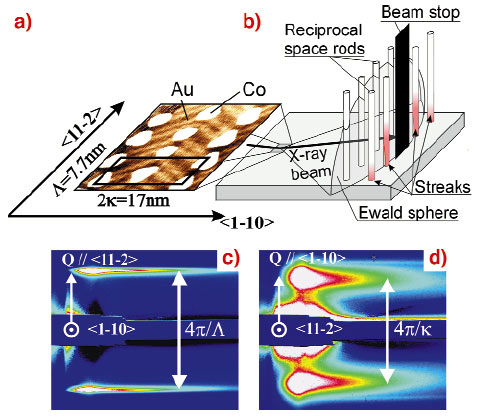
Sub-atomic-layer Co deposition on reconstructed Au(111) yields a self-organised array of parallel rows of dots, with a period around 10 nm (Figure 100) [2]. The reciprocal space of this array consists of rods perpendicular to the surface, with scattering vectors connected with the super-cell of the array. Due to the high radius of the Ewald sphere scattering patterns consist of streaks elongated perpendicular to the sample's surface. The order of dots within rows (resp. between rows) is revealed with the beam shone perpendicular (resp. parallel) to the rows (Figure 100). The order is found to be of crystalline type within rows (narrow streaks) and of liquid type between the rows (broad peak).
 |
|
Fig. 100: (a) STM view of self-organised Co/Au(111) dots (b) Sketch of the experimental geometry (c-d) GISAXS patterns for two azimuths of the X-ray beam. The sample lies vertical at the left-hand side of the patterns. |
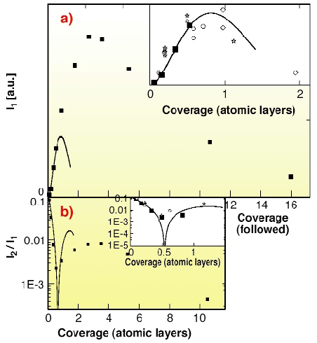
For self-organised samples, the analogy between real-time GISAXS and reflection high-energy electron diffraction (RHEED) is striking, except that GISAXS investigates dots instead of atoms, and is more suitable to quantitative analysis because of the weak interaction between X-rays and matter. To illustrate this point, Figure 101 displays the evolution during growth of the intensities I1 and (normalised) I2/I1 of the first and second order peaks of Figure 100c (second order peaks not shown on in the figure). The intensity reflects both the order of the array and the shape function of the individual dots, explaining the very different behaviours of I1 and I2/I1. In the sub-atomic-layer range the data is well reproduced by simulations based on Scanning Tunnelling Microscopy (STM) images and on a model of perfect percolation. For higher coverage percolation into a continuous film progressively occurs. The slow decay of I1 with respect to the model indicates that the percolation is imperfect. Much smaller values are also expected from STM images, revealing that a significant periodic microstructure remains buried in the film even after apparent percolation is probed by STM at the free surface.
 |
|
Fig. 101: GISAXS intensity versus Co/Au(111) coverage for first (top) and second (bottom) order streaks. Squares: experimental GISAXS (azimuth of Figure 100c) ; lines: model for perfect percolation ; symbols (insets only): intensity calculated from STM images topography. |
In conclusion GISAXS is a promising technique to investigate self-organised deposits on surfaces in real time, revealing the reciprocal space of the array's super-cell. By a quantitative peak intensity analysis, like in conventional crystallography, valuable information is deduced on dots shape, size, order and percolation.
References
[1] G. Renaud, M. Noblet, A. Barbier, C. Revenant, O. Ulrich, Y. Borensztein, R. Lazzari, J. Jupille, C. Henry, ESRF Highlights 1999, 41 (1999).
[2] B. Voigtländer, G. Meyer, N.M. Amer, Phys. Rev. B, 44, 10354 (1991).
Principal Publication and Authors
O. Fruchart (a), G. Renaud (b), A. Barbier (b,c), M. Noblet (b), O. Ulrich (b), J.-P. Deville (d), F. Scheurer (d), J. Mane-Mane (d), V. Repain (e), G. Baudot (e) and S. Rousset (e), Europhys. Lett., 63, 275-281 (2003).
(a) LLN, CNRS, Grenoble (France)
(b) CEA/DRFMC, Grenoble (France)
(c) CEA/DRECAM, Gif-sur-Yvette (France)
(d) IPCMS, Strasbourg (France)
(e) GPS-Jussieu, Paris (France)


