- Home
- Users & Science
- Scientific Documentation
- ESRF Highlights
- ESRF Highlights 2003
- X-ray Absorption and Magnetic Scattering
- Isolating Interface Magnetocrystalline Anisotropy Contributions in Magnetic Multilayers
Isolating Interface Magnetocrystalline Anisotropy Contributions in Magnetic Multilayers
Magnetic valves are used billions of times each day to read information from computer hard disks. These valves rely on a phenomenon known as giant magnetoresistance which in turn is highly sensitive to interface magnetism in magnetic multilayers. However, interface magnetism is still relatively poorly understood due largely to the lack of a suitable probe. Fortunately, recent work on beamline ID08 has shown that Soft X-ray Resonant Magnetic Scattering (SXRMS) combined with Soft X-ray Standing Waves (SXSW) offers unique new insights into interface magnetism. Fe/CeH2 multilayers provide an ideal system to demonstrate the magnetic interface sensitivity of this new probe since the magnetisation orientates perpendicular to the layers and splits up into a regular spin-up and spin-down stripe domain configuration at low temperatures. The stripe domains act, so to speak, as a magnetic diffraction grating for SXRMS [1] while the high quality of the multilayer interfaces produces standing waves allowing interface sensitivity. For transition metal/rare-earth interfaces, very little is understood about the mechanism behind this spin reorientation. On one hand, the strong interface crystalline field acts on the 4f orbital moment resulting in a single-ion anisotropy model of the spin reorientation. On the other hand, the magnetocrystalline anisotropy energy (MAE) of the transition metal 3d states could directly induce the spin reorientation, without a contribution from the 4f orbital moment, through interface 3d - 5d hybridisation. Despite the significance of these phenomena, making a distinction between the two mechanisms has proven difficult.
 |
|
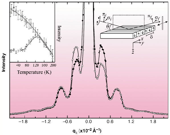
Fig. 119: SXRMS spectra measured at the Fe L3 edge using left (open circles) and right (solid circles) circularly-polarised light. The inset shows the intensity changes of the first- (open squares) and second-order (open circles) peaks as a function of temperature; the lines are guides to the eye. The experimental geometry is shown with the up and down arrows representing the stripe domains. |
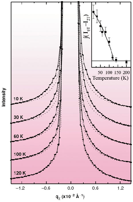
Soft X-ray resonant magnetic scattering is sensitive to the element-specific stripe domain structure of Fe and Ce through resonant transitions to the 3d and 4f unoccupied states, respectively. Figure 119 shows SXRMS spectra measured, at the Fe L3 edge, using left (open circles) and right (solid circles) circularly polarised light. The integrated intensity variation of the first- (open squares) and second-order (open circles) magnetic satellites with temperature is shown in the inset Figure 119. The different behaviour of the two features is clearly evident below 120K and it can be shown that the second-order peak is related to the spin-orbit anisotropy and therefore a probe of the magnetocrystalline anisotropy [2]. The deviation between the first and second-order magnetic Fe SXRMS contributions at 120K arises from an additional magnetic anisotropy contribution due to a Ce 4f single-ion anisotropy. Evidence for such a mechanism comes from magnetic scattering at the Ce M5 edge. Figure 120 shows SXRMS spectra recorded at the Ce M5 edge as a function of temperature (solid circles with lines); the spectra recorded after the stripes have been destroyed in a magnetic field are also shown (dashed lines). The inset of Figure 120 shows the integrated difference between the pairs of spectra for each temperature, which represents the Ce 4f perpendicular moment and MAE. The temperature dependence of the Ce 4f single-ion anisotropy contribution is noticeably different since the weak first-order magnetic Bragg peaks are only visible below 100 K (much lower than the spin reorientation temperature), as shoulders.
 |
|
Fig. 120: Temperature dependence of the Ce SXRMS spectra (solid circles with lines). The SXRMS scattering recorded after the stripes have been destroyed in an applied magnetic field of 2T is also shown for each temperature (dashed lines). The inset shows the temperature dependence of the integrated difference between the spectra recorded with and without a 2T field and represents the temperature dependence of the Ce 4f perpendicular magnetic moment and magnetic anisotropy. |
The first- and second-order resonant magnetic scattering contributions have been related to the interface and bulk regions of the Fe layers using SXSW. They are generated within the multilayer due to the long wavelength matching the multilayer periodicity. Interface sensitivity was achieved by tuning the standing-wave maxima to coincide with the interfaces. In this way, a difference in the angular dependence of the magnetic satellites demonstrated that the first- and second-order contributions probe different magnetic properties localised at the bulk Fe and interface Fe sites. At the spin reorientation, the magnetic anisotropy driving the perpendicular magnetic anisotropy is then dominated by the Fe interface MAE, which most likely arises from Ce 5d - Fe 3d hybridisation, and not the Ce 4f single-ion anisotropy.
References
[1] H. A. Dürr et. al., Science, 284, 2166 (1999).
[2] S. S. Dhesi, G. van der Laan, E. Dudzik, A. B. Shick, Phys. Rev. Lett., 87, 067201 (2001).
Principal Publication and Authors
S.S. Dhesi (a,b), H.A. Dürr (c), M. Münzenberg (d), W. Felsch (d) Phys. Rev. Lett., 90, 117204 (2003).
(a) ESRF
(b) Diamond Light Source (UK)
(c) BESSY (Germany)
(d) Universität Göttingen (Germany)


